Category
page 1Cardiac electrophysiology
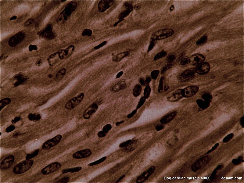
myocardium
middle layer of the heart wall, which consists of cardiac muscle

electrocardiography
thumb|Use of monitoring (medicine)|real time monitoring of the heart in an [[intensive care unit in a German hospital (2015), the monitoring screen above the patient displaying an electrocardiogram and various values of parameters of the heart like heart rate and blood pressure]]

artificial pacemaker
A pacemaker, also known as an artificial cardiac pacemaker, is an implanted medical device that generates electrical pulses delivered by electrodes to one or more of the chambers of the heart. Each pulse causes the targeted chamber(s) to contract and pump blood, thus regulating the function of the electrical conduction system of the heart.

defibrillation
Defibrillation is a treatment for life-threatening cardiac arrhythmias, specifically ventricular fibrillation (V-Fib) and non-perfusing ventricular tachycardia (V-Tach). Defibrillation delivers a dose of electric current (often called a counter-shock) to the heart. Although not fully understood, this process depolarizes a large amount of the heart muscle, ending the arrhythmia. Subsequently, the body's natural pacemaker in the sinoatrial node of the heart is able to re-establish normal sinus rhythm. A heart which is in asystole (flatline) cannot be restarted by defibrillation; it would be trea
automated external defibrillator
portable electronic device that automatically diagnoses the life-threatening cardiac arrhythmias of ventricular fibrillation and pulseless ventricular tachycardia in a patient
electrical conduction system of the heart
transmits signals generated usually by the sinoatrial node to cause contraction of the heart muscle
antiarrhythmic agent
pharmaceutical used to suppress abnormal rhythms of the heart
subendocardial branches
fibers that allow the heart's conduction system to create synchronized contractions of its ventricles
atrioventricular bundle
collection of heart muscle cells specialized for electrical conduction
implantable cardioverter-defibrillator
device implantable inside the body, able to perform cardioversion, defibrillation, and (in modern versions) pacing of the heart
QT interval
measure of the time between the start of the Q wave and the end of the T wave in the heart's electrical cycle
cardiac pacemaker
Network of cells that facilitate rhythmic heart contraction
refractory period
in physiology
bundle branch block
defect of the bundle branches or fascicles in the electrical conduction system of the heart
ST segment
the ST segment connects the QRS complex and the T wave and has a duration of 0.080 to 0.120 sec
QRS complex
combination of three of the graphical deflections seen on a typical electrocardiogram
P wave
represents atrial depolarization, which results in atrial contraction
PR interval
the period, measured in milliseconds, that extends from the beginning of the P wave (the onset of atrial depolarization) until the beginning of the QRS complex (the onset of ventricular depolarization)
cardiac action potential
biological process in the heart

T wave
represents the repolarization (or recovery) of the ventricles
Cable theory
Mathematical model of a dendrite
Reversal potential
in a biological membrane
U wave
ECG waveform thought to represent repolarization of the Purkinje fibers
Cardiac monitoring
medical diagnostic method
sinus arrhythmia
irregular heart rate caused by abnormal function of the sinoatrial node
implantable loop recorder
ILR
Einthoven's triangle
concept in electrocardiography
KCNQ1
Potassium voltage-gated channel subfamily KQT member 1 is a potassium channel protein encoded in the human by the KCNQ1 gene. Its mutation causes long QT syndrome, Kv7.1 is a voltage and lipid-gated potassium channel present in the cell membranes of cardiac tissue and in inner ear neurons among other tissues. In the cardiac cells, Kv7.1 mediates the IKs (or slow delayed rectifying K+) current that contributes to the repolarization of the cell, terminating the cardiac action potential and thereby the heart's contraction. It is a member of the KCNQ family of potassium channels.
cardiac resynchronization therapy
treatment for heart failure
ballistocardiography
Ballistocardiography (BCG) is the measurement of ballistic forces generated by the heart. The downward movement of blood through the descending aorta produces an upward recoil, moving the body upward with each heartbeat. As different parts of the aorta expand and contract, the body continues to move downward and upward in a repeating pattern. Ballistocardiography is a technique for producing a graphical representation of repetitive motions of the human body arising from the sudden ejection of blood into the great vessels with each heart beat. It is a vital sign in the 1–20 Hz frequency range w
subcutaneous implantable defibrillator
implantable medical device
transvenous pacing
medical intervention
Electrophysiology study
Medical test.
Afterdepolarization
Afterdepolarizations are abnormal depolarizations of cardiac myocytes that interrupt phase 2, phase 3, or phase 4 of the cardiac action potential in the electrical conduction system of the heart. Afterdepolarizations may lead to cardiac arrhythmias. Afterdepolarization is commonly a consequence of myocardial infarction, cardiac hypertrophy, or heart failure. It may also result from congenital mutations associated with calcium channels and sequestration.
cardiac electrophysiology
science of elucidating, diagnosing and treating the electrical activities of the heart