Category
page 1Long bones

femur
The femur (; : femurs or femora ), or thigh bone is the only bone in the thigh — the region of the lower limb between the hip and the knee. In many four-legged animals, the femur is the upper bone of the hindleg.

tibia
The tibia (; : tibiae or tibias), also known as the shinbone, shankbone or simply the shin, is the larger, stronger, and anterior (frontal) of the two bones in the leg below the knee in vertebrates (the other being the fibula, behind and to the outside of the tibia); it connects the knee with the ankle. The tibia is found on the medial side of the leg next to the fibula and closer to the median plane. The tibia is connected to the fibula by the interosseous membrane of leg, forming a type of fibrous joint called a syndesmosis with very little movement. The tibia is named for the flute tibia. I

humerus
The humerus (; : humeri) is a long bone in the arm that runs from the shoulder to the elbow. It connects the scapula and the two bones of the lower arm, the radius and ulna, and consists of three sections. The humeral upper extremity consists of a rounded head, a narrow neck, and two short processes (tubercles, sometimes called tuberosities). The shaft is cylindrical in its upper portion, and more prismatic below. The lower extremity consists of 2 epicondyles, 2 processes (trochlea and capitulum), and 3 fossae (radial fossa, coronoid fossa, and olecranon fossa). As well as its true anatomical
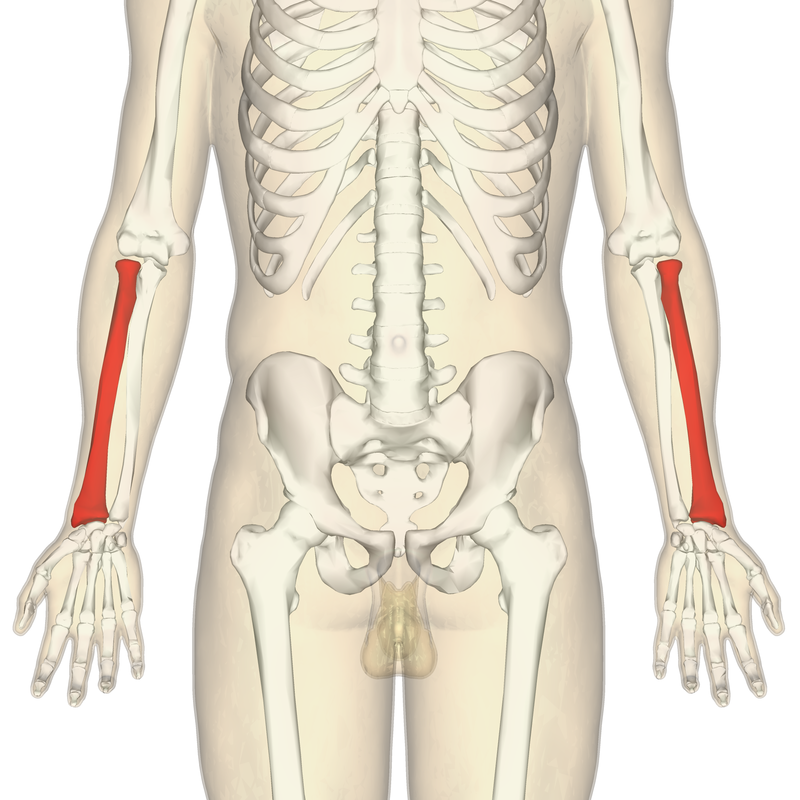
radius
one of the two long bones of the forearm

fibula
alt=3D Medical Animation still shot of Fibula structure|thumb|260x260px|3D medical animation still shot of fibula structure
The fibula (: fibulae or fibulas) or calf bone is a leg bone on the lateral side of the tibia, to which it is connected above and below. It is the smaller of the two bones and, in proportion to its length, the most slender of all the long bones. Its upper extremity is small, placed toward the back of the head of the tibia, below the knee joint and excluded from the formation of this joint. Its lower extremity inclines a little forward, so as to be on a plane anterior to t

ulna
thumb|Right ulna, lateral aspect. (After Piersol.)
thumb|Right ulnar bone, medial aspect. (After Piersol.)
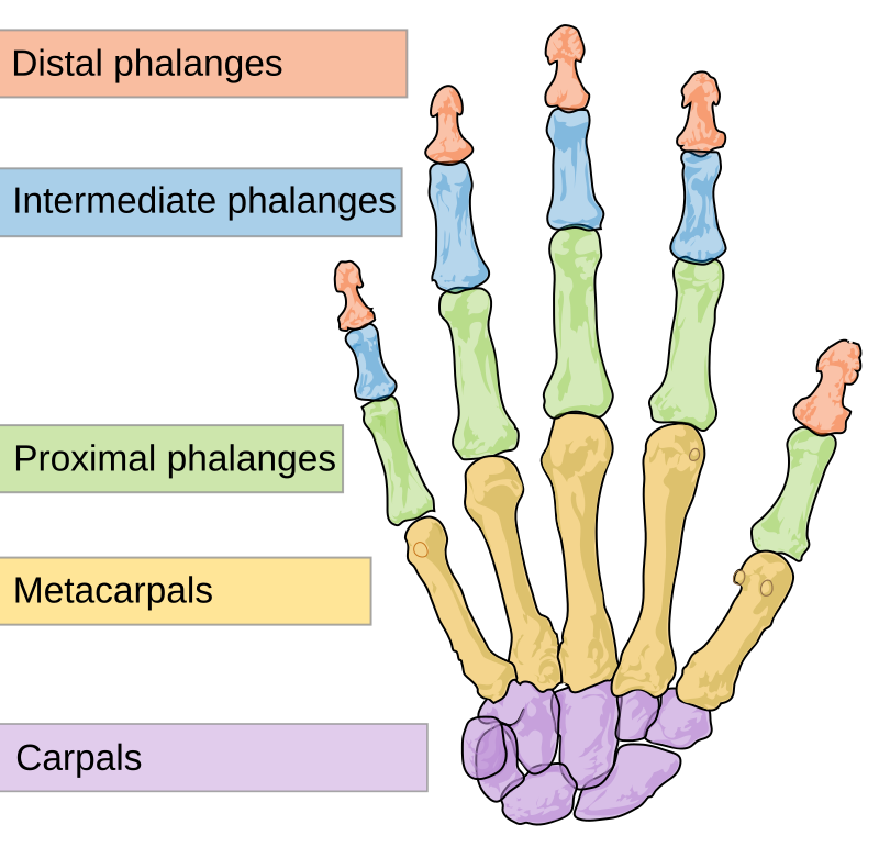
phalanx
digital bone in the hands and feet of most vertebrates

metacarpal bones
bones of hand

epiphysis
An epiphysis (; : epiphyses) is one of the rounded ends or tips of a long bone that ossify from one or more secondary centers of ossification. Between the epiphysis and diaphysis (the long midsection of the long bone) lies the metaphysis, including the epiphyseal plate (growth plate). During formation of the secondary ossification center, vascular canals (epiphysial canals) stemming from the perichondrium invade the epiphysis, supplying nutrients to the developing secondary centers of ossification. At the joint, the epiphysis is covered with articular cartilage; below that covering is a zone s
long bone
bone that is longer than it is wide
diaphysis
The diaphysis (: diaphyses) is the main or midsection (shaft) of a long bone. It is made up of cortical bone and usually contains bone marrow and adipose tissue (fat).

metaphysis
The metaphysis (: metaphyses) is the neck portion of a long bone between the epiphysis and the diaphysis. It contains the growth plate, the part of the bone that grows during childhood, and as it grows it ossifies near the diaphysis and the epiphyses. The metaphysis contains a diverse population of cells including mesenchymal stem cells, which give rise to bone and fat cells, as well as hematopoietic stem cells which give rise to a variety of blood cells as well as bone-destroying cells called osteoclasts. Thus the metaphysis contains a highly metabolic set of tissues including trabecular (spo