Category
page 1Cerebrum

cerebrum
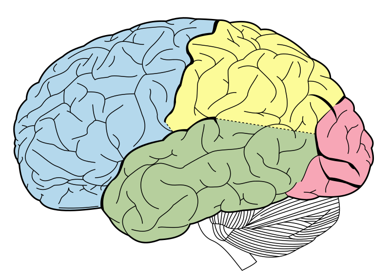
The cerebrum (: cerebra), telencephalon or endbrain is the largest part of the brain, containing the cerebral cortex (of the two cerebral hemispheres) as well as several subcortical structures, including the hippocampus, basal ganglia, and olfactory bulb. In the human brain, the cerebrum is the uppermost region of the central nervous system. The cerebrum develops prenatally from the forebrain (prosencephalon). In mammals, the dorsal telencephalon, or pallium, develops into the cerebral cortex, and the ventral telencephalon, or subpallium, becomes the basal ganglia. The cerebrum is also divided
occipital lobe
Part of the brain that is located at the back of the head and is responsible for visual perception. That includes colour, form and motion. Damage to the occipital lobe can cause difficulty with locating objects in environment.
frontal lobe
part of the brain that, because of how large it is, it's responsible for many functions, such as motor control, executive functions, language production, emotional regulation, working memory, and personality
parietal lobe
part of the brain responsible for sensory input and some language processing

cerebral hemisphere
half of the cerebrum
temporal lobe
part of the brain responsible for processing auditory information and encoding of memory. The temporal lobe also plays a role in processing affect/emotions, language, and certain aspects of visual perception.

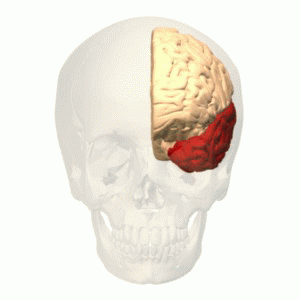
Broca's area
region of the brain in the frontal lobe - in the dominant hemisphere - that aids speech production

prosencephalon
In the anatomy of the brain of vertebrates, the forebrain or prosencephalon is the rostral (forward-most) portion of the brain. The forebrain controls body temperature, reproductive functions, eating, sleeping, and the display of emotions.
cerebral edema
human disease

striatum
The striatum (: striata) or corpus striatum is a cluster of interconnected nuclei that make up the largest structure of the subcortical basal ganglia. The striatum is a critical component of the motor and reward systems; receives glutamatergic and dopaminergic inputs from different sources; and serves as the primary input to the rest of the basal ganglia.
olfactory bulb
neural structure of the vertebrate forebrain involved in olfaction, which sends olfactory information to be further processed in the amygdala, the orbitofrontal cortex and the hippocampus where it plays a role in emotion, memory and learning
Brodmann area
Region of the cerebral cortex defined according to its cytoarchitecture.
optic chiasm
optical part of brain

gyrus
thumb|300px|Gray's Anatomy|Gray's FIG. 726 – Lateral surface of left [[cerebral hemisphere, viewed from the side]]
thumb|300px|Gray's Anatomy|Gray's Fig. 727 – Medial surface of left cerebral hemisphere
Claustrum
The claustrum (Latin, meaning "to close" or "to shut") is a thin sheet of neurons and supporting glial cells in the brain that connects to the cerebral cortex and subcortical regions including the amygdala, hippocampus and thalamus. It is located between the insular cortex laterally and the putamen medially, encased by the extreme and external capsules respectively. Blood to the claustrum is supplied by the middle cerebral artery. It is considered to be the most densely connected structure in the brain, and thus hypothesized to allow for the integration of various cortical inputs such as visio
fornix of the brain
a part of limbic syst
lateralization of brain function
tendency for cognitive processes to be specialized to one side of the brain or the other
longitudinal cerebral fissure
Deep Groove that separates the two-halves of the Brain
high altitude cerebral edema
medical condition
cerebral peduncle
band of neurons, resembling a stalk, which connect varied parts of the brain

split-brain procedure
Split-brain or callosal syndrome is a type of disconnection syndrome when the corpus callosum connecting the two hemispheres of the brain is severed to some degree. It is an association of symptoms produced by disruption of, or interference with, the connection between the hemispheres of the brain. The surgical operation to produce this condition (corpus callosotomy) involves transection of the corpus callosum, and is usually a last resort to treat refractory epilepsy. Initially, partial callosotomies are performed; if this operation does not succeed, a complete callosotomy is performed to mit
optic (nerve) tract
nerve fiber originating from the optic chiasm
Two-streams hypothesis#Dorsal stream
model of the neural processing of vision and hearing
cortical homunculus
distorted model of the human body based on areas and proportions of the brain dedicated to motor or sensory functions for different body parts
external capsule
part of the brain
precentral sulcus
part of the human brain
extreme capsule
part of white matter in the brain
septum pellucidum
thin membrane between the lateral ventricles of the brain
preoccipital notch
part of the human brain

leukoaraiosis
thumb|Axial T2 FLAIR sequence MR image of a middle-aged man with leukoaraiosis.
thumb|right|MRI image: Leukoaraiosis in a 90-year-old patient with cerebral atrophy.
thumb|Head CT showing periventricular white matter lesions.
Leukoaraiosis is a particular abnormal change in appearance of white matter near the lateral ventricles. It is often seen in aged individuals, but sometimes in young adults. On MRI, leukoaraiosis changes appear as white matter hyperintensities (WMHs) in T2 FLAIR images. On CT scans, leukoaraiosis appears as hypodense periventricular white-matter lesions.
pallium
layers of nerve cells on the surface of cerebral hemispheres of chordate animals
Operculum
cover the insula as the opercula of insula
Orbital gyri
Brain regions
stria terminalis
Band of fibres along the thalamus
collateral fissure
brain structure
archicortex
The archicortex, or archipallium, is the phylogenetically second oldest region of the brain's cerebral cortex (the oldest is the paleocortex). In older species, such as fish, the archipallium makes up most of the cerebrum. Amphibians develop an archipallium and paleopallium.
cingulum
structure in human brain
paracentral sulcus
sulcus of the brain
postcentral sulcus
Anatomical furrow of the brain
paleocortex
In anatomy of animals, the paleocortex, or paleopallium, is a region within the telencephalon in the vertebrate brain. This type of cortical tissue consists of three cortical laminae (layers of neuronal cell bodies). In comparison, the neocortex has six layers and the archicortex has three or four layers. Because the number of laminae that compose a type of cortical tissue seems to be directly proportional to both the information-processing capabilities of that tissue and its phylogenetic age, paleocortex is thought to be an intermediate between the archicortex (or archipallium) and the neocor
Cortical minicolumn
structure in the brain
Interpeduncular fossa
brain segment
septal nuclei
limbic system brain structure
Functional specialization
theory that regions of the brain are specialized for functions