Category
page 1Pelvis

uterus
The uterus (from Latin uterus, : uteri or uteruses) or womb () is the organ in the reproductive system of most female mammals, including humans, that accommodates the embryonic and fetal development of one or more fertilized eggs until birth. The uterus is a hormone-responsive sex organ that contains glands in its lining that secrete uterine milk for embryonic nourishment. (The term uterus is also applied to analogous structures in some non-mammalian animals.)

Vulva
In mammals, the vulva (: vulvas or vulvae) comprises mostly external, visible structures of the female genitalia leading into the interior of the female reproductive tract. For humans, it includes the mons pubis, labia majora, labia minora, clitoris, vestibule, urinary meatus, vaginal introitus, hymen, and openings of the vestibular glands (Bartholin's and Skene's). The folds of the outer and inner labia provide a double layer of protection for the vagina (which leads to the uterus). While the vagina is a separate part of the anatomy, colloquially the term has often been used synonymously with

urinary bladder
The bladder () is a hollow organ in humans and other vertebrates that stores urine from the kidneys. In placental mammals, urine enters the bladder via the ureters and exits via the urethra during urination. In humans, the bladder is a distensible organ that sits on the pelvic floor. The typical adult human bladder will hold between 300 and (10 and ) before the urge to empty occurs, but can hold considerably more.
.jpg)
ovary
The ovary () is a gonad in the female reproductive system that produces ova; when released, an ovum travels through the fallopian tube/oviduct into the uterus. There is an ovary on the left and the right side of the body. The ovaries are endocrine glands, secreting various hormones that play a role in the menstrual cycle and fertility. The ovary progresses through many stages beginning in the prenatal period through menopause.

pelvis
thumb|250px|The same human pelvis, front imaged by X-ray (top), [[magnetic resonance imaging (middle), and three-dimensional computed tomography (bottom)]]
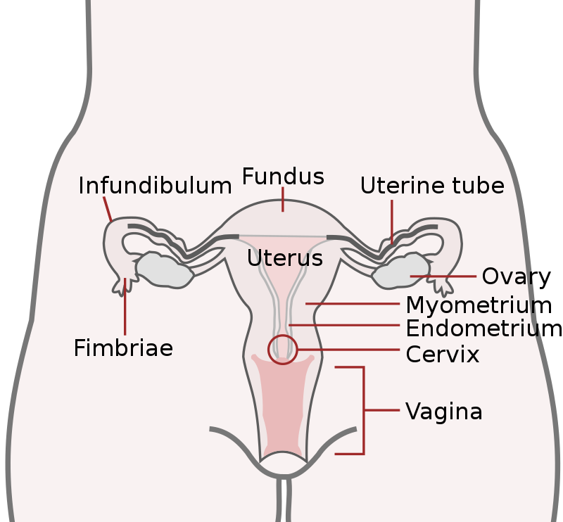
fallopian tube
part of the female reproductive organs

foreskin
In male human anatomy, the foreskin, also known as the prepuce (), is the double-layered fold of skin, mucosal and muscular tissue at the distal end of the human penis that covers the glans and the urinary meatus. The foreskin is attached to the glans by an elastic band of tissue, known as the frenulum. The outer skin of the foreskin meets with the inner preputial mucosa at the area of the mucocutaneous junction. The foreskin is mobile, fairly stretchable and sustains the glans in a moist environment. Except for humans, a similar structure known as a penile sheath appears in the male sexual or

perineum
The perineum (: perineums or perinea) in placental mammals is the space between the anus and the genitals. The human perineum is between the anus and scrotum in the male or between the anus and vulva in the female. The perineum is the region of the body between the pubic symphysis (pubic arch) and the coccyx (tail bone), including the perineal body and surrounding structures. The perineal raphe is visible and pronounced to varying degrees.

mons pubis
rounded mass of fatty tissue found over the pubic symphysis

corpus luteum
temporary endocrine structure in ovaries producing progesterone, estradiol and inhibin A; the remains of an ovarian follicle that has released an egg

endometrium
The endometrium is the inner epithelial layer, along with its mucous membrane, of the mammalian uterus. It has a basal layer and a functional layer: the basal layer contains stem cells which regenerate the functional layer. The functional layer thickens and then is shed during menstruation in humans and some other mammals, including other apes, Old World monkeys, some species of bat, the elephant shrew and the Cairo spiny mouse. In most other mammals, the endometrium is reabsorbed in the estrous cycle. During pregnancy, the glands and blood vessels in the endometrium further increase in size a
pubic symphysis
cartilaginous joint that sits between and joins the left and right superior rami of the pubic bones
myometrium
The myometrium is the middle layer of the uterine wall, consisting mainly of uterine smooth muscle cells (also called uterine myocytes) but also of supporting stromal and vascular tissue. Its main function is to induce uterine contractions.
levator ani
broad, thin muscle, situated on either side of the pelvis
hip replacement surgery
surgery replacing hip joint with prosthetic implant
Greg pouch
anatomical location between the rectum and uterus, sometimes traditionally called the "cul-de-sac"
pelvic cavity
body cavity bounded by the bones of the pelvis
sacroiliac joint
joint
pelvic fracture
fractures of the hip bone
corpus albicans
structure derived from the corpus luteum
pelvic floor dysfunction
medical condition

Pelvic thrust
Thrusting motion of the pelvic region
elongated labia
natural elongation of the labia minora
Iliac fascia
a fascia
epilploic appendages of colon
one of several small pouches of fat on the peritoneum along the colon and rectum
gluteal sulcus
fold separating the thigh from the buttock
Uterosacral ligament
major ligaments of uterus
pelvimetry
Pelvimetry is the measurement of the female pelvis. It can theoretically identify cephalo-pelvic disproportion, which is when the capacity of the pelvis is inadequate to allow the fetus to negotiate the birth canal. However, clinical evidence indicate that all pregnant women should be allowed a trial of labor regardless of pelvimetry results.
Protrusio acetabuli
human disease
obturator canal
passageway
sacrococcygeal symphysis
joint in the pelvis
obstetrical dilemma
hypothesis about human childbirth
symphysis pubis dysfunction
medical condition
pelvic tilt
orientation of the pelvis in respect to the thighbones and the rest of the body
pelvic binder
medical device used to compress and stabilize the pelvis after pelvic fracture
pelvic inlet
part of the pelvis
obturator fascia
Rectoprostatic fascia
peritoneoperineal fascia
Risser sign
indirect measure of skeletal maturity
pelvic fascia