Category
page 1Skull

skull
The skull, or cranium, is typically a bony enclosure around the brain of a vertebrate. In some fish and amphibians, the skull is of cartilage. The skull is at the head end of the vertebrate.
cochlea
thumb|3D model of cochlea and semicircular canals
The cochlea is the part of the inner ear involved in hearing. It is a spiral-shaped cavity in the bony labyrinth, in humans making 2.75 turns around its axis, the modiolus. A core component of the cochlea is the organ of Corti, the sensory organ of hearing, which is distributed along the partition separating the fluid chambers in the coiled tapered tube of the cochlea.
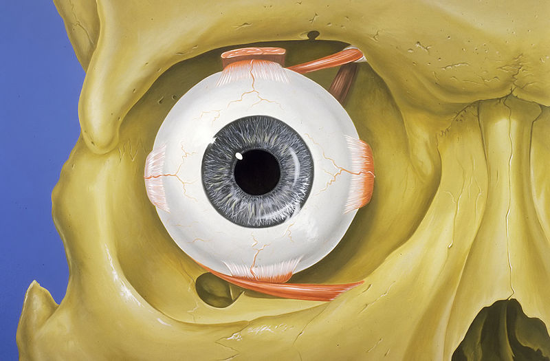
orbit
cavity or socket of the skull in which the eye and its appendages are situated
fontanelle
A fontanelle (or fontanel) (colloquially, soft spot) is an anatomical feature of the infant human skull comprising soft membranous gaps (sutures) between the cranial bones that make up the calvaria of a fetus or an infant. Fontanelles allow for stretching and deformation of the neurocranium both during birth and later as the brain expands faster than the surrounding bone can grow. Premature complete ossification of the sutures is called craniosynostosis.
temporomandibular joint
Joints connecting the jawbone to the skull
artificial cranial deformation
Form of body alteration

posterior nasal apertures
thumb|right|The choanae (internal nostrils) of a cat, indicated by the dashed lines and bounded by the vomer (blue gray) and the [[palatine bone (orange)]]
calvaria
upper part of the neurocranium that covers the cranial cavity containing the brain
zygomatic arch
cheek bone
base of skull
inferior area of the skull, composed of the endocranium and lower parts of the skull roof
bregma
The bregma is the anatomical point on the skull at which the coronal suture is intersected perpendicularly by the sagittal suture.
sagittal crest
ridge of bone along the top of a skull
lambdoid suture
dense, fibrous connective tissue joint on the posterior aspect of the skull
coronal suture
connective tissue joint of the skull
cranial cavity
space inside the skull formed by eight cranial bones know as the neurocranium
vertex
the upper surface of the head

headbutt
thumb|Caricature of capoeira carioca from Rio, using cocada headbutt.
temporal fossa
Shallow depression on the side of the human skull
infratemporal fossa
cavity that is part of the skull

Pterion peor
thumb|Sphenoparietal suture highlighted in red
The pterion is the region where the frontal, parietal, temporal, and sphenoid bones join. It is located on the side of the skull, just behind the temple. It is also considered to be the weakest part of the skull, which makes it clinically significant, as if there is a fracture around the pterion it could be accompanied by an epidural hematoma.
asterion
bone meeting point in the skull
lambda
structure in the skull

frontal suture
Midline joint in the frontal bone of the forehead
Starchild skull
archaeological find

posterior cranial fossa
part of the cranial cavity, located between the foramen magnum and tentorium cerebelli

carotid canal
Passage in the skull's temporal bone
Skull cup
Cup made from a skull
squamosal suture
cranial suture between the temporal squama and the parietal bone
mastoid cell
air-filled cavities in the temporal bone
Diploë
Diploë ( or ) is the spongy cancellous bone separating the inner and outer layers of the cortical bone of the skull. It is a subclass of trabecular bone.
jugular process
Part of the human skull
styloid process of temporal bone
part of the temporal bone
fibrous joint
fixed joints between bones held together by dense, fibrous tissue
Anterior fontanelle
Fontanelle in human skull
occipitomastoid suture
cranial suture between the occipital bone and the temporal bone
sphenosquamosal suture
cranial suture
incisive foramen
funnel-shaped opening in the bone of the oral hard palate immediately behind the incisor teeth where blood vessels and nerves pass
anterior cranial fossa
part of skull housing the projecting frontal lobes of the brain
mastoid antrum
air space in the petrous portion of the temporal bone
middle cranial fossa
separated from the posterior fossa by the clivus and the petrous crest
Frontoethmoidal suture
suture between the ethmoid bone and the frontal bone
mastoid part of the temporal bone
Posterior part of the temporal bone
sphenozygomatic suture
cranial suture between the sphenoid bone and the zygomatic bone
sphenofrontal suture
cranial suture between the sphenoid bone and the frontal bone
zygomaticotemporal suture
Rigid joint between zygomatic bone (cheekbone) and temporal bone
zygomaticofrontal suture
cranial suture
Sphenoethmoidal suture
anatomical feature of the human skull
sphenoparietal suture
cranial suture between the sphenoid bone and the parietal bone
sphenopetrosal fissure
suture between sphenoid bone and petrous part of temporal bone
occipital bun
prominent bulge of the occipital bone at the back of the skull
Posterior fontanelle
gap between bones in the human skull
mastoid foramen
large hole in the posterior border of the temporal bone
Skull roof
roofing bones of the skull
mastoidectomy
A mastoidectomy is a procedure performed to remove the mastoid air cells near the middle ear. The procedure is part of the treatment for mastoiditis, chronic suppurative otitis media or cholesteatoma. Additionally, it is sometimes performed as part of other procedures, such as cochlear implants, or to access the middle ear.
pterygomaxillary fissure
anatomical fissure human skull
postorbital bar
straight or arched rod of bone that forms the posterior rim of the eye opening (orbit) in amniote skulls
stephanion
The point where the upper temporal line cuts the coronal suture is named the stephanion.
rabbit punch
blow to the back of the head or to the base of the skull
chondrocranium
The chondrocranium (or cartilaginous neurocranium) is the primitive cartilaginous skeletal structure of the fetal skull that grows to envelop the rapidly growing embryonic brain.