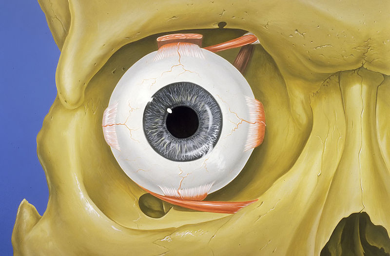
File:Eye_orbit_anatomy_anterior2.jpg · Wikimedia Commons · See Wikimedia Commons
orbit
Sign in to saveAlso known as ocular orbit, orbital cavity, eye socket, eye-socket
cavity or socket of the skull in which the eye and its appendages are situated
Key facts
- Latin
- orbita
- Mesh
- D009915
- Ta98
- A02.1.00.067 , A01.2.01.010
via Wikipedia infobox
Research
147,052 papers- Anatomy of the orbit.Neuroimaging clinics of North America · 1998
- Inflammatory sinus diseases affecting the orbit.Otolaryngologic clinics of North America · 1993
- Application of image-guidance to surgery of the orbit.Otolaryngologic clinics of North America · 2005
- Embryologic and Fetal Development of the Human Orbit.Ophthalmic plastic and reconstructive surgery · 2018
- MR-Eye: High-Resolution Microscopy Coil MRI for the Assessment of the Orbit and Periorbital Structures, Part 2: Clinical Applications.AJNR. American journal of neuroradiology · 2021
via PubMed
Wikidata facts
- Part of
- face
- Has part
- Inferior orbital fissure
- Image
- Eye orbit anterior.jpg
Show 4 more facts
- Commons category
- Orbit (anatomy)
- Commons gallery
- Orbit (anatomy)
- NCI Thesaurus ID
- C12347
- topic has template
- Template:Orbital anatomy
Sources (6)
via Wikidata · CC0
~9 min read
Encyclopedic overview
In vertebrate anatomy, the orbit is the cavity or socket/hole of the skull in which the eye and its appendages are situated. "Orbit" can refer to the bony socket, or it can also be used to imply the contents. In the adult human, the volume of the orbit is about 28 millilitres (0.99 imp fl oz; 0.95 US fl oz), of which the eye occupies 6.5 ml (0.23 imp fl oz; 0.22 US fl oz). The orbital contents comprise the eye, the orbital and retrobulbar fascia, extraocular muscles, cranial nerves II, III, IV, V, and VI, blood vessels, fat, the lacrimal gland with its sac and duct, the eyelids, medial and lateral palpebral ligaments, cheek ligaments, the suspensory ligament, septum, ciliary ganglion and short ciliary nerves.
Structure
Excerpted from Wikipedia’s “orbit” article, available under the CC BY-SA 4.0 licence.