Category
page 1Visual system

eye
An eye is a sensory organ that allows an organism to perceive visual information. It detects light and converts it into electro-chemical impulses in neurons (neurones). It is part of an organism's visual system.

retina
The retina (; or retinas) is the innermost, light-sensitive layer of tissue of the eye of most vertebrates and some molluscs. The optics of the eye create a focused two-dimensional image of the visual world on the retina, which then processes that image within the retina and sends nerve impulses along the optic nerve to the visual cortex to create visual perception. The retina serves a function which is in many ways analogous to that of the film or image sensor in a camera.

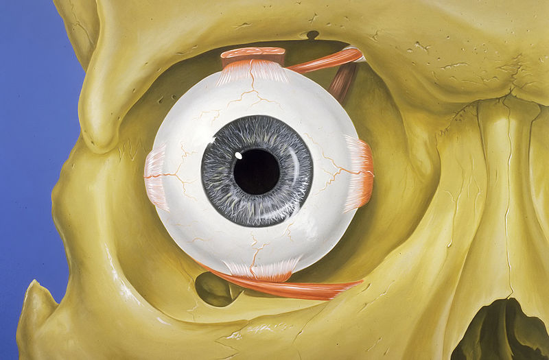
human eye
mammalian eye; part of the visual organ of the human body

optic nerve
second cranial nerve, which connects the eyes to the brain

George Wald
American biologist, biochemist, physiologist and Nobel laureate (1906–1997)

visual system
system of body parts responsible for sight

oculomotor nerve
cranial nerve III, for eye movements
orbit
cavity or socket of the skull in which the eye and its appendages are situated

prosopagnosia
Prosopagnosia, also known as face blindness, is a cognitive disorder of face perception in which the ability to recognize familiar faces, including one's own face (self-recognition), is impaired, while other aspects of visual processing (e.g., object discrimination) and intellectual functioning (e.g., decision-making) remain intact. The term originally referred to a condition following acute brain damage (acquired prosopagnosia), but a congenital or developmental form of the disorder also exists, with a prevalence of 2–2.5%.

choroid
The choroid, also known as the choroidea or choroid coat, is a part of the uvea, the vascular layer of the eye. It contains connective tissues, and lies between the retina and the sclera. The human choroid is thickest at the far extreme rear of the eye (at 0.2 mm), while in the outlying areas it narrows to 0.1 mm. The choroid provides oxygen and nourishment to the outer layers of the retina. Along with the ciliary body and iris, the choroid forms the uveal tract.

phosphene
An artist's representation of how some people may see phosphenes by retinal stimulation|alt=artistic representation of phosphenes|thumb
A phosphene is the phenomenon of seeing light without light entering the eye. The word phosphene comes from the Greek words phos (light) and phainein (to show). Phosphenes that are induced by movement or sound may be associated with optic neuritis.
visual field
total area or space visible in a person's peripheral vision with the eye looking straight forward
optic chiasm
optical part of brain
retinal ganglion cell
type of neuron located near the inner surface (ganglion cell layer) of the retina of the eye
achromatopsia
Achromatopsia, also known as rod monochromacy, is a medical syndrome that exhibits symptoms relating to five conditions, most notably monochromacy. Historically, the name referred to monochromacy in general, but now typically refers only to an autosomal recessive congenital color vision condition. The term is also used to describe cerebral achromatopsia, though monochromacy is usually the only common symptom. The conditions include: monochromatic color blindness, poor visual acuity, and day-blindness. The syndrome is also present in an incomplete form that exhibits milder symptoms, including r

trichromacy
thumb|310px|Close-up of a trichromatic in-line shadow mask [[CRT display, which creates most visible colors through combinations and different levels of the three primary colors: red, green and blue]]

saccade
thumb|Trace of saccades of the human eye on a face while scanning
thumb|200px |Saccades during observation of a picture on a computer screen
lateral geniculate nucleus
Relay Centre in Thalamus for Optic Reflexes
superior colliculus
structure in the mammalian midbrain

emmetropia
Emmetropia is the state of vision in which a faraway object at infinity is in sharp focus with the ciliary muscle in a relaxed state. That condition of the normal eye is achieved when the refractive power of the cornea and eye lens and the axial length of the eye balance out, which focuses rays exactly on the retina, resulting in perfectly sharp distance vision. A human eye in a state of emmetropia requires no corrective lenses for distance; the vision scores well on a visual acuity test (such as an eye chart test).
optic (nerve) tract
nerve fiber originating from the optic chiasm
visual agnosia
impairment in recognition of visually presented objects
visual phototransduction
sensory transduction of the visual system
Two-streams hypothesis#Dorsal stream
model of the neural processing of vision and hearing
corneal endothelium
a single layer of cells on the inner surface of the cornea
intrinsically photosensitive retinal ganglion cell
neuron in the retina of the mammalian eye
Grassmann's law
Perception of color mixtures
optic radiation
neural pathway in the visual system
fusiform face area
part of the human visual system that is specialized for facial recognition
pretectal area
structure in the midbrain which mediates responses to ambient light
redout
A redout occurs when the body experiences a negative g-force sufficient to cause a blood flow from the lower parts of the body to the head. It is the inverse effect of a greyout, where blood flows away from the head to the lower parts of the body. Usually, a redout will only ever be experienced by aircraft pilots, as planes are the most common devices that allow such negative g-forces to be exerted. Redouts are potentially dangerous and can cause retinal damage and hemorrhagic stroke.
optokinetic reflex
eye phenomenon, normal nystagmus produced by looking at objects moving across the field of vision
greyout
thumb|right|300px|Simulated stages of a greyout.
A greyout is a transient loss of vision characterized by a perceived dimming of light and color, sometimes accompanied by a loss of peripheral vision. It is a precursor to fainting or a blackout and is caused by hypoxia (low brain oxygen level), often due to a loss of blood pressure.
smooth pursuit
eye movement used when tracking objects
retinotopy
thumb|Retinotopic maps with explanation|right|350px
parasol cell
cell type

phantom eye syndrome
condition of pain in a lost eye
Optic cup
finding on fundoscopy
midget cell
type of retinal ganglion cell