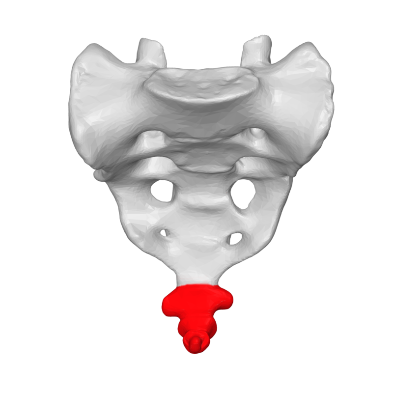
File:Coccyx_-_anterior_view02.png · Wikimedia Commons · See Wikimedia Commons
coccyx
Sign in to saveAlso known as tailbone, coxis
The coccyx (: coccyges or coccyxes), commonly referred to as the tailbone, is the final segment of the vertebral column in all apes, and analogous structures in certain other mammals such as horses. In tailless primates (e.g. humans and other great apes) since Nacholapithecus (a Miocene hominoid), the coccyx is the remnant of a vestigial tail. In animals with bony tails, it is known as tailhead or dock, in bird anatomy as tailfan. It comprises three to five separate or fused coccygeal vertebrae below the sacrum, attached to the sacrum by a fibrocartilaginous joint, the sacrococcygeal symphysis
The coccyx, commonly called the tailbone, is the final segment of the spine located at the base of the backbone below the sacrum. In humans and other great apes, it is a remnant of what was once a functional tail, and it consists of three to five vertebrae that are either separate or fused together.
AI-generated from the Wikipedia summary — may contain errors.
Key facts
- Bone.Name
- Coccyx
- Bone.Latin
- os coccygis
- Bone.Image
- Coccyx - anterior view02.png
- Bone.Caption
- The coccyx
- Bone.Image2
- Illu vertebral column.svg
- Bone.image2_class
- skin-invert
- Bone.Caption2
- The coccyx is the final bone in the vertebral column that surrounds the spinal cord.
via Wikipedia infobox
Research
1,872 papers- Coccyx pain in women after childbirth.ReviewEnfermeria clinica · 2019Márquez-Carrasco ÁM, García-García E, Aragúndez-Marcos MPDOI: 10.1016/j.enfcli.2019.01.005
- Coccyx pain.Tidsskrift for den Norske laegeforening : tidsskrift for praktisk medicin, ny raekke · 2026Knobloch RG, Kalstad AM, Finsen VDOI: 10.4045/tidsskr.25.0421
- Coccyx fractures treated with intranasal calcitonin.ReviewPain physician · 2014Foye PM, Shupper P, Wendel I
- COCCYGODYNIA.ReviewActa clinica Croatica · 2023Elezović N, Stojanović Stipić S, Perković M et al.DOI: 10.20471/acc.2023.62.s4.14
- Sacrum and Coccyx Shape Changes During Pregnancy and After Delivery.Annals of biomedical engineering · 2024Martin LC, Routzong MR, Moalli PA et al.DOI: 10.1007/s10439-023-03375-y
- Cement or Calcitonin for Coccyx Fractures.Balkan medical journal · 2021Foye PM, Massey HR, Koger TJDOI: 10.4274/balkanmedj.galenos.2020.2020.8.34
- Neonatal coccyx reverse curvature deformity: A case report.Asian journal of surgery · 2023Mao X, Pan S, Yang X et al.DOI: 10.1016/j.asjsur.2023.06.134
- Disorders of the coccyx and sacrococcygeal joint: Etiology, diagnosis, and management strategies.ReviewThe American journal of medicine · 2026Carayannopoulos NL, Montaquila NS, Lewis AM et al.DOI: 10.1016/j.amjmed.2025.10.038
via PubMed
Wikidata facts
- Image
- Gray100.png
Show 3 more facts
- Commons category
- Coccyx
- different from
- Kostrč
- NCI Thesaurus ID
- C12696
Sources (7)
via Wikidata · CC0
~8 min read
Encyclopedic overview
16 sectionsContents
- Structure
- Surfaces
- Borders
- Apex
- Coccygeal fossa
- Extensor coccygis
- Sacrococcygeal and intercoccygeal joints
- Attachments
- Function
- Clinical significance
- Etymology
- Additional images
- See also
- References
- Further reading
- External links
The coccyx (: coccyges or coccyxes), commonly referred to as the tailbone, is the final segment of the vertebral column in all apes, and analogous structures in certain other mammals such as horses. In tailless primates (e.g. humans and other great apes) since Nacholapithecus (a Miocene hominoid), the coccyx is the remnant of a vestigial tail. In animals with bony tails, it is known as tailhead or dock, in bird anatomy as tailfan. It comprises three to five separate or fused coccygeal vertebrae below the sacrum, attached to the sacrum by a fibrocartilaginous joint, the sacrococcygeal symphysis, which permits limited movement between the sacrum and the coccyx.
== Structure == 250px|thumb|A coccyx with four vertebrae below the sacrum The coccyx is formed of three, four or five rudimentary vertebrae. It articulates superiorly with the sacrum. In each of the first three segments may be traced a rudimentary body and articular and transverse processes; the last piece (sometimes the third) is a mere nodule of bone. The transverse processes are most prominent and noticeable on the first coccygeal segment. All the segments lack pedicles, laminae and spinous processes. The first segment is the largest; it resembles the lowest sacral vertebra, and often exists as a separate piece; the remaining ones diminish in size caudally.
Excerpted from Wikipedia’s “coccyx” article, available under the CC BY-SA 4.0 licence.
Gallery (6)
Available in 62 languages
- Español
- Français
- Deutsch
- 中文
- 日本語
- Русский
- Português
- Italiano
- العربية
- हिन्दी
- Afrikaans
- Atikamekw
- Azerbaijani
- Bahasa Indonesia
- Bangla
- Bashkir
- Basque
- Belarusian
Show 43 more
- Bosnian
- Breton
- Bulgarian
- Catalan
- cdo
- Central Kurdish
- Chuvash
- Czech
- Danish
- Esperanto
- Estonian
- Finnish
- Galician
- Greek
- Hebrew
- Ido
- Irish
- Kazakh
- Latvian
- Limburgish
- Lithuanian
- Luxembourgish
- Malay
- Nederlands
- Newari
- Norwegian
- Polski
- Romanian
- Serbian
- Sicilian
- simple
- Slovak
- Slovenian
- Svenska
- Tamil
- Türkçe
- Ukrainian
- Welsh
- Western Mari
- zh_min_nan
- zh_yue
- فارسی
- 한국어
via Wikidata sitelinks · CC0