Category
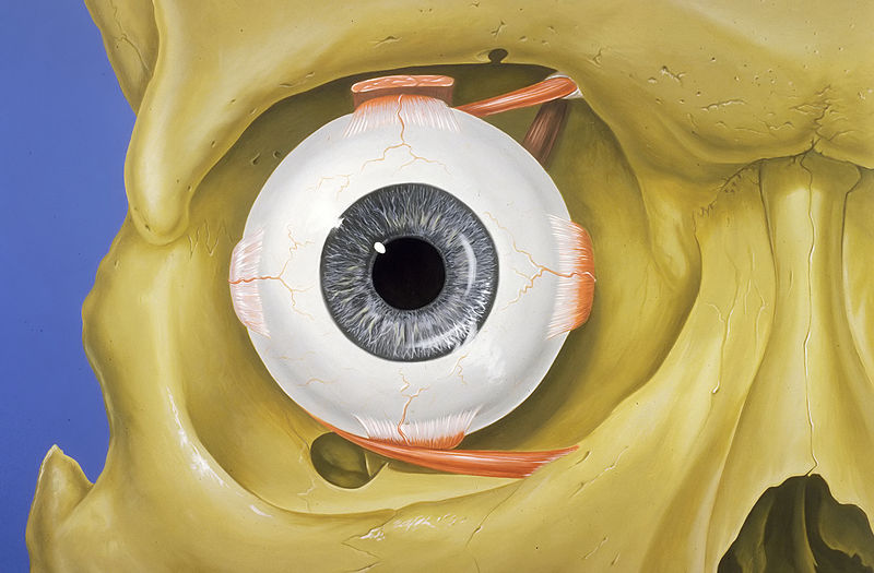
page 1Human eye anatomy

retina
The retina (; or retinas) is the innermost, light-sensitive layer of tissue of the eye of most vertebrates and some molluscs. The optics of the eye create a focused two-dimensional image of the visual world on the retina, which then processes that image within the retina and sends nerve impulses along the optic nerve to the visual cortex to create visual perception. The retina serves a function which is in many ways analogous to that of the film or image sensor in a camera.

eyelid
thumb|Blood vessels of the eyelids, front view

cornea
The cornea is the transparent front part of the eye which covers the iris, pupil, and anterior chamber. Along with the anterior chamber and lens, the cornea refracts light, accounting for approximately two-thirds of the eye's total optical power. In humans, the refractive power of the cornea is approximately 43 dioptres. The cornea can be reshaped by surgical procedures such as LASIK.

lens
transparent structure in the eye

sclerasdcbn
The sclera, also known as the white of the eye or, in older literature, as the tunica albuginea oculi, is the opaque, fibrous, protective outer layer of the eye containing mainly collagen and some crucial elastic fiber.

cone cell
photo sensitive cells that detect color
orbit
cavity or socket of the skull in which the eye and its appendages are situated

rod cell
photoreceptor cells that can function in lower light better than cone cells.

vitreous humour
clear gel that fills the space between the lens and the retina of the eyeball

choroid
The choroid, also known as the choroidea or choroid coat, is a part of the uvea, the vascular layer of the eye. It contains connective tissues, and lies between the retina and the sclera. The human choroid is thickest at the far extreme rear of the eye (at 0.2 mm), while in the outlying areas it narrows to 0.1 mm. The choroid provides oxygen and nourishment to the outer layers of the retina. Along with the ciliary body and iris, the choroid forms the uveal tract.

conjunctiva
thumb|Image of a human eye showing the blood vessels of the bulbar conjunctiva
thumb|Hyperaemia of the superficial bulbar conjunctiva blood vessels

macula of retina
The macula (), in full macula lutea, is an oval-shaped pigmented area in the center of the retina of the human eye and in other animals. The macula in humans has a diameter of around and is subdivided into the umbo, foveola, foveal avascular zone, fovea, parafovea, and perifovea areas.
photoreceptor cell
specialized type of cell found in the retina that is capable of visual phototransduction
epicanthic fold
fold on upper eye lid
aqueous humour
transparent, watery, fluid similar to plasma, but containing low protein concentrations, secreted from the ciliary epithelium
lacrimal gland
paired, almond-shaped exocrine gland, one for each eye, that secretes the aqueous layer of the tear film
ciliary body
part of an eye
uvea
The uvea (; derived from meaning "grape"), also called the uveal layer, uveal coat, uveal tract, vascular tunic or vascular layer, is the pigmented middle layer of the three concentric layers that make up an eye, precisely between the inner retina and the outer fibrous layer composed of the sclera and cornea.
ciliary muscle
eye muscle used for focusing
fovea centralis
anatomical structure
anterior chamber of the eye
space in the eye between the cornea and lens which contains the aqueous humor
Schlemm's canal
lymphatic-like vessel in the eye
meibomian glands
set of exocrine glands, along the rims of the eyelid
retinal ganglion cell
type of neuron located near the inner surface (ganglion cell layer) of the retina of the eye
lacrimal apparatus
physiological system containing the orbital structures for tear production and drainage
extraocular muscle
seven extrinsic muscles of the human eye
nasolacrimal duct
carries tears from the lacrimal sac of the eye into the nasal cavity
inferior oblique muscle
part of the eye
optic disc
part of the eye
superior rectus muscle
extraocular muscle that elevates the eye
levator palpebrae superioris muscle
Muscle in orbit that elevates upper eyelid
superior oblique muscle
part of the eye
medial rectus muscle
extraocular muscle that rotates the eye medially
retinal pigment epithelium
Layer of cells in the eye
lacrimal sac
upper, dilated end of the nasolacrimal duct
inferior rectus muscle
muscle in the orbit
retina amacrine cell
cell type
zonule of Zinn
part of the eye
lateral rectus muscle
muscle on lateral side of the eye
posterior chamber of eyeball
region of the eyeball between the iris and lens
corneal limbus
the border of the cornea and the sclera
Descemet's membrane
layer of the cornea, basal lamina of the corneal endothelium
fundus
concave interior of the eye
Bowman's membrane
layer in the cornea of the eye
Dua's layer
layer of the human cornea
trabecular meshwork
area of tissue in the eye
retina bipolar cell
type of neuron
lacrimal canaliculi
small channels in each eyelid that drain lacrimal fluid

crystallins
thumb|Crystal structure of Duck Delta 1 Crystallin, based on the Protein Data Bank|PDB file 1HY0.
In anatomy, a crystallin is a water-soluble structural protein found in the lens and the cornea of the eye accounting for the transparency of the structure. It has also been identified in other places such as the heart, and in aggressive breast cancer tumors.
The physical origins of eye lens transparency and its relationship to cataract are an active area of research. Since it has been shown that lens injury may promote nerve regeneration,
crystallin has been an area of neural research. So far, it
anterior segment of eyeball
front third of the eye
corneal endothelium
a single layer of cells on the inner surface of the cornea
tarsus
connective tissue in the eyelid
retina horizontal cell
cell type
anulus tendineus communis
muscle arise from annular tendon is medial rectus,superior rectus,inferior rectus and lateral rectus
fibrous tunic of eyeball
part of a human eye
intrinsically photosensitive retinal ganglion cell
neuron in the retina of the mammalian eye
Bruch's membrane
membrane in the eye
ora serrata
where rod and cone terminate
ciliary process
folded layers in the anterior of the eye
lacrimal caruncle
small, reddish-yellowish, globular nodule at the inner corner (the medial angle) of the eye