Category
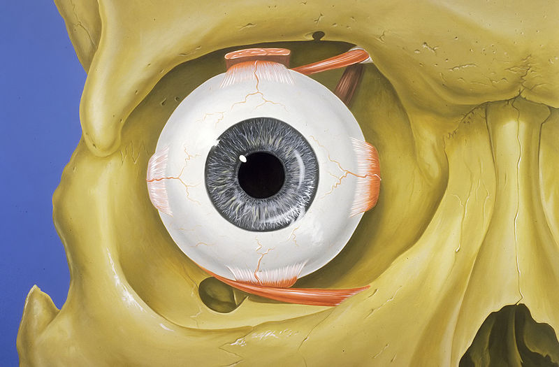
page 1Ophthalmology
%20PHIL%204284%20lores.jpg)
cataract
A cataract is a cloudy area in the lens of the eye that impairs vision. Cataracts often develop slowly and can affect one or both eyes. Symptoms may include faded colours, blurry or double vision, halos around light, trouble with bright lights, and difficulty seeing at night. This may result in difficulty driving, reading and recognizing faces. Poor vision caused by cataracts may also result in an increased risk of falling and depression. In 2020 Cataracts caused 39.6% of all cases of blindness and 28.3% of visual impairment worldwide. Cataracts remain the single most common cause of global bl

ophthalmology
Ophthalmology (, ) is the branch of medicine that deals with the diagnosis, treatment, and surgery of eye diseases and disorders.

eyelid
thumb|Blood vessels of the eyelids, front view

human eye
mammalian eye; part of the visual organ of the human body

optic nerve
second cranial nerve, which connects the eyes to the brain

sunglasses
thumb|upright=1.2|Wearing sunglasses under direct sunlight: Large lenses offer good protection, but broad temple arms are also needed against "stray light" from the sides.
orbit
cavity or socket of the skull in which the eye and its appendages are situated

trochlear nerve
4th cranial nerve

abducens nerve
cranial nerve controlling the movement of the lateral rectus muscle
visual acuity
clarity of vision

accommodation
focusing ability of eye

miosis
Miosis, or myosis (), is excessive constriction of the pupil. The opposite condition, mydriasis, is the dilation of the pupil. Anisocoria is the condition of one pupil being more dilated than the other.

mydriasis
Mydriasis is the dilation of the pupil, usually having a non-physiological cause, or sometimes a physiological pupillary response. Non-physiological causes of mydriasis include disease, trauma, or the use of certain types of drugs. It may also be of unknown cause.
lacrimal gland
paired, almond-shaped exocrine gland, one for each eye, that secretes the aqueous layer of the tear film
retinal detachment
human disease

rheum
thumb|right|Rheum from a cat's eyes
Rheum (; from Greek: ῥεῦμα rheuma 'a flowing, rheum') is a thin mucus naturally discharged from the eyes, nose, or mouth, often during sleep (contrast with mucopurulent discharge). Rheum dries and gathers as a crust in the corners of the eyes or the mouth, on the eyelids, or under the nose. It is formed by a combination of mucus (in the case of the eyes, consisting of mucin discharged from the cornea or the conjunctiva), nasal mucus, blood cells, skin cells, or dust.
intraocular pressure
fluid pressure inside the eye

ketorolac
Ketorolac, sold under the brand name Toradol, Acular and Sprix, among others, is a nonsteroidal anti-inflammatory drug (NSAID) used to treat pain. Specifically it is recommended for moderate to severe pain. Recommended duration of treatment is less than six days, and in Switzerland not more than seven days (parenterally two days). It is used by mouth, by nose, by injection into a vein or muscle, and as eye drops. Effects begin within an hour and last for up to eight hours. Ketorolac also has antipyretic (fever-reducing) properties.
visual field
total area or space visible in a person's peripheral vision with the eye looking straight forward
field of view
extent of the observable world seen at any given moment
Nd:YAG laser
type of laser
intraocular lens
lens implanted in the eye to treat cataracts or myopia
eye surgery
medical specialty
ocular prosthesis
type of craniofacial prosthesis

eyepatch
An eyepatch is a small patch that is worn in front of one eye. It may be a cloth patch attached around the head by an elastic band or by a string, an adhesive bandage, or a plastic device which is clipped to a pair of glasses. It is often worn by people to cover a lost, infected, or injured eye, but it also has a therapeutic use in children for the treatment of amblyopia. Eyepatches used to block light while sleeping are referred to as a sleep mask.
pupillary light reflex
reflex controlling the diameter of the pupil in response to the intensity of light
Bates method
alternative eyesight improvement therapy
lacrimal sac
upper, dilated end of the nasolacrimal duct
entoptic phenomenon
visual effect whose source is within the eye itself
corrective lens
transmissive optical device worn on the eye to improve visual perception
rose bengal
tetrachloro-tetraiodo-fluorescein used as stain
oculocardiac reflex
Pulse rate connected with eye muscles
corneal endothelium
a single layer of cells on the inner surface of the cornea
herpes simplex virus keratitis
keratitis of the cornea that has material basis in herpes simplex type infection
Lisch nodule
pigmented hamartomatous nodular aggregate of dendritic melanocytes affecting the iris
epiretinal membrane
disease of the eye in response to changes in the vitreous humor or more rarely, diabetes
ophthalmic zoster
human disease

keratometer
thumb|An eye doctor examining a patient with a keratometer
thumb|Typical presentations of keratoconus as detected through a keratometer
thumb|Shin Nippon Nvision K-5001 Refkeratometer
A keratometer, also known as an ophthalmometer, is a diagnostic instrument for measuring the curvature of the anterior surface of the cornea, particularly for assessing the extent and axis of astigmatism. It was invented by the German physiologist Hermann von Helmholtz in 1851, although an earlier model was developed in 1796 by Jesse Ramsden and Everard Home.

neuro-ophthalmology
Neuro-ophthalmology is an academically-oriented subspecialty that merges the fields of neurology and ophthalmology, often dealing with complex systemic diseases that have manifestations in the visual system. Neuro-ophthalmologists initially complete a residency in either neurology or ophthalmology, then do a fellowship in the complementary field. Since diagnostic studies can be normal in patients with significant neuro-ophthalmic disease, a detailed medical history and physical exam is essential, and neuro-ophthalmologists often spend a significant amount of time with their patients.

ocular dominance
tendency of the brain to prefer visual input from one eye over the other

blue field entoptic phenomenon
tiny bright dots moving quickly in the visual field

solar viewer
type of eye-wear used to view the sun
Landolt C
Optotype
eye black
undereye makeup worn by sports players

subluxation
A subluxation is an incomplete or partial dislocation of a joint or organ. According to the World Health Organization, a subluxation is a "significant structural displacement" and is therefore visible on static imaging studies, such as X-rays. Unlike real subluxations, the pseudoscientific concept of a chiropractic "vertebral subluxation" may or may not be visible on x-rays.
high-energy visible light
effects of blue light
Bell's phenomenon
reflex of the eye
Keratoscope
A keratoscope, sometimes known as ''Placido's disk'', is an ophthalmic instrument used to assess the shape of the anterior surface of the cornea. A series of concentric rings is projected onto the cornea and their reflection viewed by the examiner through a small hole in the centre of the disk. A regular-shaped cornea should show equally spaced symmetric reflections. If the patient is suffering from astigmatism or from a corneal dystrophy, the rings will be distorted.
osteo-odonto-keratoprosthesis
Osteo-odonto-keratoprosthesis (OOKP), also known as "tooth in eye" surgery, is a medical procedure to restore vision in the most severe cases of corneal and ocular surface patients. It includes removal of a tooth from the patient or a donor.
phoropter
thumb|right|A phoropter can measure refractive error to determine an individual's spectacle lens prescription during an eye examination.
thumb|right|Side of a phoropter that faces the patient
Monoyer chart
eye chart used to measure visual acuity
eyeglass prescription
order written by an optometrist or ophthalmologist, specifying parameters of corrective lenses for a particular patient
congenital cataract
disorder of lens
accommodation reflex
reflex action of the eye, in response to focusing on a near object, then looking at a distant object
corneal keratocyte
cell type
tebentafusp
Tebentafusp, sold under the brand name Kimmtrak, is an anti-cancer medication used to treat uveal melanoma (eye cancer). Tebentafusp is a bispecific gp100 peptide-HLA-directed CD3 T cell engager. Tebentafusp is given by intravenous infusion.
Susana Marcos Celestino
Spanish scientist, physicist and physiologist
Microkeratome
A microkeratome is a precision surgical instrument with an oscillating blade designed for creating the corneal flap in LASIK or ALK surgery. The normal human cornea varies from around 500 to 600 μm in thickness; and in the LASIK procedure, the microkeratome creates an 83 to 200 μm thick flap. The microkeratome uses an oscillating blade system, which has a blade that oscillates horizontally as the blade travels vertically for a precise cut. This piece of equipment is used all around the world to cut the cornea flap. The microkeratome is also used in Descemet's stripping automated endothelial ke
gland of Zeis
Oil glands on the margin of the eyelid
Autorefractor
thumb|A United States Navy optometrist technician using an autorefractor during a humanitarian assistance project in [[Nicaragua in 2008]]
An autorefractor or automated refractor is a computer-controlled machine used during an eye examination to provide an objective measurement of a person's refractive error and prescription for glasses or contact lenses. This is achieved by measuring how light is changed as it enters a person's eye.