Category
page 1Skin anatomy

melanin
thumb|Micrograph of melanin pigment (light refracting granular material—center of image) in a pigmented [[melanoma]]
thumb|Micrograph of the epidermis, with melanin labeled at left

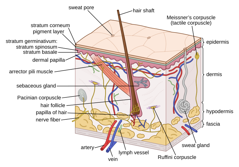
epidermis
The epidermis is the outermost of the three major layers that constitute the skin, the inner layers being the dermis and hypodermis. The epidermal layer provides a barrier to infection from environmental pathogens and regulates the amount of water released from the body into the atmosphere through transepidermal water loss.

keratins
thumb|300px|Microscopy of keratin filaments inside cells
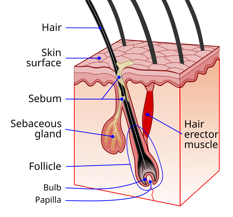
sebaceous gland
microscopic exocrine gland in the skin that secretes an oily or waxy matter, called sebum, to lubricate and waterproof the skin and hair of mammals
sweat gland
small tubular structures of the skin that produce sweat; a type of exocrine gland, which are glands that produce and secrete substances onto an epithelial surface by way of a duct

dermis
The dermis or corium is a layer of skin between the epidermis (with which it makes up the cutis) and subcutaneous tissues, that primarily consists of dense irregular connective tissue and cushions the body from stress and strain. It is divided into two layers, the superficial area adjacent to the epidermis called the papillary region and a deep thicker area known as the reticular dermis. The dermis is tightly connected to the epidermis through a basement membrane. Structural components of the dermis are collagen, elastic fibers, and extrafibrillar matrix. It also contains mechanoreceptors that

melanocyte
Melanocytes are melanin-producing neural crest-derived cells located in the bottom layer (the stratum basale) of the skin's epidermis, the middle layer of the eye (the uvea), the inner ear, vaginal epithelium, meninges, bones, and heart found in many mammals and birds. Melanin is a dark pigment primarily responsible for skin color. Once synthesized, melanin is contained in special organelles called melanosomes which can be transported to nearby keratinocytes to induce pigmentation. Thus darker skin tones have more melanosomes present than lighter skin tones. Functionally, melanin serves as pro

epicanthic fold
fold on upper eye lid
subcutaneous tissue
lowermost layer of the integumentary system in vertebrates

keratinocyte
thumb|Micrograph of keratinocytes, Stratum basale|basal cells and [[melanocytes in the epidermis]] thumb|Keratinocytes (stained green) in the skin of a mouse
dermatome
area of the skin which is supplied by a spinal nerve
Merkel cell
type of cutaneous mechanoreceptor cel
Langerhans cell
cell type
melanosome
thumb|7× speed timelapse video of fish melanophores responding to 200 uM adrenaline; the melanosomes retreat to the center of the star-shaped melanophore cells.
thumb|Fish and frog melanophores are cells that can change colour by dispersing or aggregating pigment-containing melanosomes.
hemidesmosome
Hemidesmosomes are very small stud-like structures found in keratinocytes of the epidermis of skin that attach to the extracellular matrix. They are similar in form to desmosomes when visualized by electron microscopy; however, desmosomes attach to adjacent cells. Hemidesmosomes are also comparable to focal adhesions, as they both attach cells to the extracellular matrix. Instead of desmogleins and desmocollins in the extracellular space, hemidesmosomes utilize integrins. Hemidesmosomes are found in epithelial cells connecting the basal epithelial cells to the basement membrane. Hemidesmosomes
basal lamina
a thin sheet of proteoglycans and glycoproteins, especially laminin, secreted by cells as an extracellular matrix.
stratum corneum
outermost layer of the epidermis
Blaschko's lines
lines on skin, believed to trace the migration of embryonic cells, invisible under normal conditions.
stratum basale
deepest layer of the five layers of the epidermis

Dewlap
thumb|right|A mastiff with a dewlap, seen connecting from the neck to the lower jaw.
A dewlap is a longitudinal flap of skin or similar flesh that hangs beneath the lower jaw or neck of many vertebrates. More loosely, it can be various similar structures in the neck area, such as those caused by a double chin or the submandibular vocal sac of a frog. More generally, it can be any hanging mass of skin, such as a fold of loose skin on an elderly person's neck, or the wattle of a bird. Dewlaps can be considered as a caruncle, defined as "a small, fleshy excrescence that is a normal part of an ani
apocrine sweat gland
mostly limited to the axilla (armpits) and perianal areas in humans; they are not significant for cooling in humans, but are the sole effective sweat glands in hoofed animals, such as the camels, donkeys, horses, and cattle
eccrine sweat gland
gland distributed almost all over the human body
stratum lucidum
a thin, clear layer of skin cells
stratum spinosum
layer of the epidermis
stratum granulosum
layer of cells in the epidermis
periorbital puffiness
appearance of swelling in the tissues around the eyes

skin appendage
skin-associated structure from skin of vertebrates
gland of Zeis
Oil glands on the margin of the eyelid
nevus cell
cell type
nasolabial fold
two skin folds in the face
follicle
small spherical group of cells containing a cavity
striated cutaneous muscle
subcutaneous tissue
inframammary fold
anatomical line below the breasts
rhabdite
Rhabdites (from Greek, rhabdos, rod) are rodlike structures in the cells of the epidermis or underlying parenchyma in certain turbellarians, and in the epidermis of nemerteans. They are discharged in mucous secretions. They are a defensive mechanism, which dissolve in water, and they are distasteful to most animals who would prey on rhabditid worms. In nemerteans, rhabdites form mucus on which the animals glide.
corneocyte
Corneocytes are terminally differentiated keratinocytes and compose most of the stratum corneum, the outermost layer of the epidermis. They are regularly replaced through desquamation and renewal from lower epidermal layers and are essential for its function as a skin barrier.
Lamina lucida
layer of the skin
panniculus adiposus
fatty layer of the subcutaneous tissue
lamellar granule
secretory organelle
Acid mantle
Capa protectora de la piel humana