Category
page 1Human cells

red blood cell
most common type of blood cell

white blood cell
type of cells of the immunological system

platelet
Platelets or thrombocytes () are a part of blood whose function (along with the coagulation factors) is to react to bleeding from blood vessel injury by clumping to form a blood clot. Platelets have no cell nucleus; they are fragments of cytoplasm from megakaryocytes which reside in bone marrow or lung tissue, and then enter the circulation. Platelets are found only in mammals, whereas in other vertebrates (e.g. birds, amphibians), thrombocytes circulate as intact mononuclear cells.

T-lymphocytes
type of lymphocyte

monocyte
Monocytes are a type of leukocyte or white blood cell. They are the largest type of leukocyte in the blood and can differentiate into macrophages and monocyte-derived dendritic cells. As a part of the vertebrate innate immune system monocytes also influence adaptive immune responses and exert tissue repair functions. There are at least three subclasses of monocytes in human blood based on their phenotypic receptors.

macrophage
Macrophages (; abbreviated Mφ, MΦ or MP) are a type of white blood cell of the innate immune system that engulf and digest pathogens, such as cancer cells, microbes, cellular debris and foreign substances, which do not have proteins that are specific to healthy body cells on their surface. This self-protection method can be contrasted with that employed by Natural Killer cells. This process of engulfment and digestion is called phagocytosis; it acts to defend the host against infection and injury.

neutrophil
Neutrophils are a type of phagocytic white blood cell and part of innate immunity. More specifically, they form the most abundant type of granulocytes and make up 40% to 70% of all white blood cells in humans. Their functions vary in different animals. In humans they participate in processes such as sterile inflammation, tissue repair, and cancer, and exhibit coordinated collective behavior. They are also known as neutrocytes, heterophils or polymorphonuclear leukocytes.

B-cell
type of white blood cell

eosinophil
Eosinophils, sometimes called eosinophiles or, less commonly, acidophils, are a variety of white blood cells and one of the immune system components responsible for combating multicellular parasites and certain infections in vertebrates. Along with mast cells and basophils, they also control mechanisms associated with allergy and asthma. They are granulocytes that develop during hematopoiesis in the bone marrow before migrating into blood, after which they are terminally differentiated and do not multiply.

basophil
Basophils are a type of white blood cell. Basophils are the least common type of granulocyte, representing about 0.5% to 1% of circulating white blood cells. They are the largest type of granulocyte. They are responsible for inflammatory reactions during immune response, as well as in the formation of acute and chronic allergic diseases, including anaphylaxis, asthma, atopic dermatitis and hay fever. They also produce compounds that coordinate immune responses, including histamine and serotonin that induce inflammation, and heparin that prevents blood clotting, although there are less than tha

cone cell
photo sensitive cells that detect color
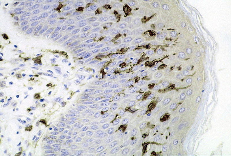
dendritic cell
specialized cells of the hematopoietic system with branch-like extensions

mast cell
granulated cell found in almost all tissues

rod cell
photoreceptor cells that can function in lower light better than cone cells.
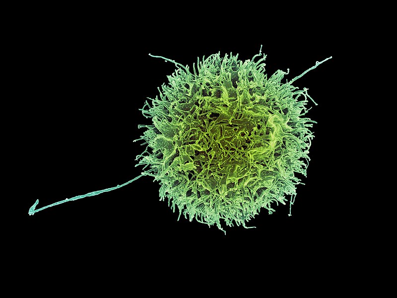
natural killer cell
type of cytotoxic lymphocyte

astrocyte
Astrocytes (from Ancient Greek , , "star" and , , "cavity", "cell"), also known collectively as astroglia, are characteristic star-shaped glial cells in the brain and spinal cord. They perform many functions, including biochemical control of endothelial cells that form the blood–brain barrier, provision of nutrients to the nervous tissue, maintenance of extracellular ion balance, regulation of cerebral blood flow, and a role in the repair and scarring process of the brain and spinal cord following infection and traumatic injuries. The proportion of astrocytes in the brain is not well defined;

melanocyte
Melanocytes are melanin-producing neural crest-derived cells located in the bottom layer (the stratum basale) of the skin's epidermis, the middle layer of the eye (the uvea), the inner ear, vaginal epithelium, meninges, bones, and heart found in many mammals and birds. Melanin is a dark pigment primarily responsible for skin color. Once synthesized, melanin is contained in special organelles called melanosomes which can be transported to nearby keratinocytes to induce pigmentation. Thus darker skin tones have more melanosomes present than lighter skin tones. Functionally, melanin serves as pro

adipocyte
Adipocytes, also known as lipocytes and fat cells, are the cells that primarily compose adipose tissue, specialized in storing energy as fat. Adipocytes are derived from mesenchymal stem cells which give rise to adipocytes through adipogenesis. In cell culture, adipocyte progenitors can also form osteoblasts, myocytes and other cell types.
plasma cell
white blood cell that secretes large volumes of antibodies
T helper cell
type of immune cell

osteoblast
Osteoblasts (from the Greek combining forms for "bone", ὀστέο-, osteo- and βλαστάνω, blastanō "germinate") are cells with a single nucleus that synthesize bone. However, in the process of bone formation, osteoblasts function in groups of connected cells. Individual cells cannot make bone. A group of organized osteoblasts together with the bone made by a unit of cells is usually called the osteon.
hepatocyte
A hepatocyte is a cell of the main parenchymal tissue of the liver. Hepatocytes make up 80% of the liver's mass.
These cells are involved in:
Protein synthesis
Protein storage
Transformation of carbohydrates
Synthesis of cholesterol, bile salts and phospholipids
Detoxification, modification, and excretion of exogenous and endogenous substances
Initiation of formation and secretion of bile

osteoclast
An osteoclast () is a type of bone cell that removes bone tissue.
This function is critical in the maintenance, repair, and remodeling of bones of the vertebral skeleton.
The osteoclast disassembles and digests the composite of hydrated protein and mineral at a molecular level by secreting acid and a collagenase, a process known as bone resorption.
This process also helps regulate the level of blood calcium.
An odontoclast (/odon·to·clast/; o-don´to-klast) is an osteoclast associated with the absorption of the roots of deciduous teeth.

megakaryocyte
A megakaryocyte () is a large bone marrow cell with a lobated nucleus that produces blood platelets (thrombocytes), which are necessary for normal clotting. In humans, megakaryocytes usually account for 1 out of 10,000 bone marrow cells, but can increase in number nearly 10-fold during the course of certain diseases. Owing to variations in combining forms and spelling, synonyms include megalokaryocyte and megacaryocyte.

reticulocyte
In hematology, reticulocytes are immature red blood cells (RBCs). In the process of erythropoiesis (red blood cell formation), reticulocytes develop and mature in the bone marrow and then circulate for about a day in the blood stream before developing into mature red blood cells. Like mature red blood cells, in mammals, reticulocytes do not have a cell nucleus. They are called reticulocytes because of a reticular (mesh-like) network of ribosomal RNA that becomes visible under a microscope with certain stains such as new methylene blue and Romanowsky stain.
goblet cell
cell type
Leydig cell
steroid-producing cells in the interstitial tissue of the testis
Sertoli cell
cell type found in testis
osteocyte
An osteocyte, an oblate-shaped type of bone cell with dendritic processes, is the most commonly found cell in mature bone. It can live as long as the organism itself. The adult human body has about 42 billion of them. Osteocytes do not divide and have an average half life of 25 years. They are derived from osteoprogenitor cells, some of which differentiate into active osteoblasts (which may further differentiate to osteocytes). Osteoblasts/osteocytes develop in mesenchyme.
cytotoxic T cell
cell
parietal cell
epithelial cell that secrete hydrochloric acid and intrinsic factor

keratinocyte
thumb|Micrograph of keratinocytes, Stratum basale|basal cells and [[melanocytes in the epidermis]] thumb|Keratinocytes (stained green) in the skin of a mouse
Kupffer cell
macrophages located in the liver
cell-mediated immunity
immune response that does not involve antibodies
retinal ganglion cell
type of neuron located near the inner surface (ganglion cell layer) of the retina of the eye
sensory neuron
nerve cell that converts environmental stimuli into corresponding internal stimuli

podocyte
Podocytes are cells in Bowman's capsule in the kidneys that wrap around capillaries of the glomerulus. Podocytes make up the epithelial lining of Bowman's capsule, the third layer through which filtration of blood takes place. Bowman's capsule filters the blood, retaining large molecules such as proteins while smaller molecules such as water, salts, and sugars are filtered as the first step in the formation of urine. Although various viscera have epithelial layers, the name visceral epithelial cells usually refers specifically to podocytes, which are specialized epithelial cells that reside in

pericyte
Pericytes (formerly called Rouget cells) are multi-functional mural cells that adhere to the external surface of the endothelial cells that form the endothelium of capillaries and other microvessels. Pericytes are embedded in the basement membrane of blood capillaries, where they communicate with endothelial cells by means of both direct physical contact and paracrine signaling. The morphology, distribution, density and molecular fingerprints of pericytes vary between organs and vascular beds. Pericytes help in the maintenance of homeostatic and hemostatic functions in the brain, where one of
erythroblast
cell type; precursor of erythrocytes
hair cell
auditory nerve cells
regulatory T cell
type of cell
chromaffin cell
cells that store epinephrine secretory vesicles
memory B cell
cell type
liver stellate cell
perisinusoidal cells of the liver, located in the space of Disse between hepatocytes and sinusoidal endothelial cells
Paneth cell
Anti-microbial epithelial cell of the small intestine
retina amacrine cell
cell type
memory T cell
cell type
Betz cell
giant pyramidal neurons of the primary motor cortex
parafollicular cell
neuroendocrine cells in the thyroid gland which secrete calcitonin
basophilia
Basophilia is the condition of having greater than 200 basophils/μL in the venous blood. Basophils are the least numerous of the myelogenous cells, and it is rare for their numbers to be abnormally high without changes to other blood components. Rather, basophilia is most often coupled with other white blood cell conditions such as eosinophilia, high levels of eosinophils in the blood. Basophils are easily identifiable by a blue coloration of the granules within each cell, marking them as granulocytes, in addition to segmented nuclei.

ameloblast
Ameloblasts are cells present only during tooth development that deposit tooth enamel, which is the hard outermost layer of the tooth forming the surface of the crown.
club cell
cell type
juxtaglomerular cell
cell type
gastric chief cell
type of gastric gland cell
pneumocyte
cell type
natural killer T cell
group of T cells that share properties of both T cells and natural killer (NK) cells
list of distinct cell types in the adult human body
Wikimedia list article
retina bipolar cell
type of neuron
enterochromaffin cell
cell type
microfold cell
cell type, lineage of epithelial cells present in mucosal tissue