Category
page 1Organelles

nucleus
membrane-bounded organelle of eukaryotic cells in which chromosomes are housed and replicated
organelle
An organelle is a specialized subunit, within a biological cell, that has a specific function. The name organelle comes from the idea that these structures are parts of cells, as organs are to the body, hence organelle, the suffix -elle being a diminutive. Organelles are either separately enclosed within their own lipid bilayers (also called membrane-bound organelles) or are spatially distinct functional units without a surrounding lipid bilayer (non-membrane bounded organelles). Although most organelles are functional units within cells, some functional units that extend outside of cells are
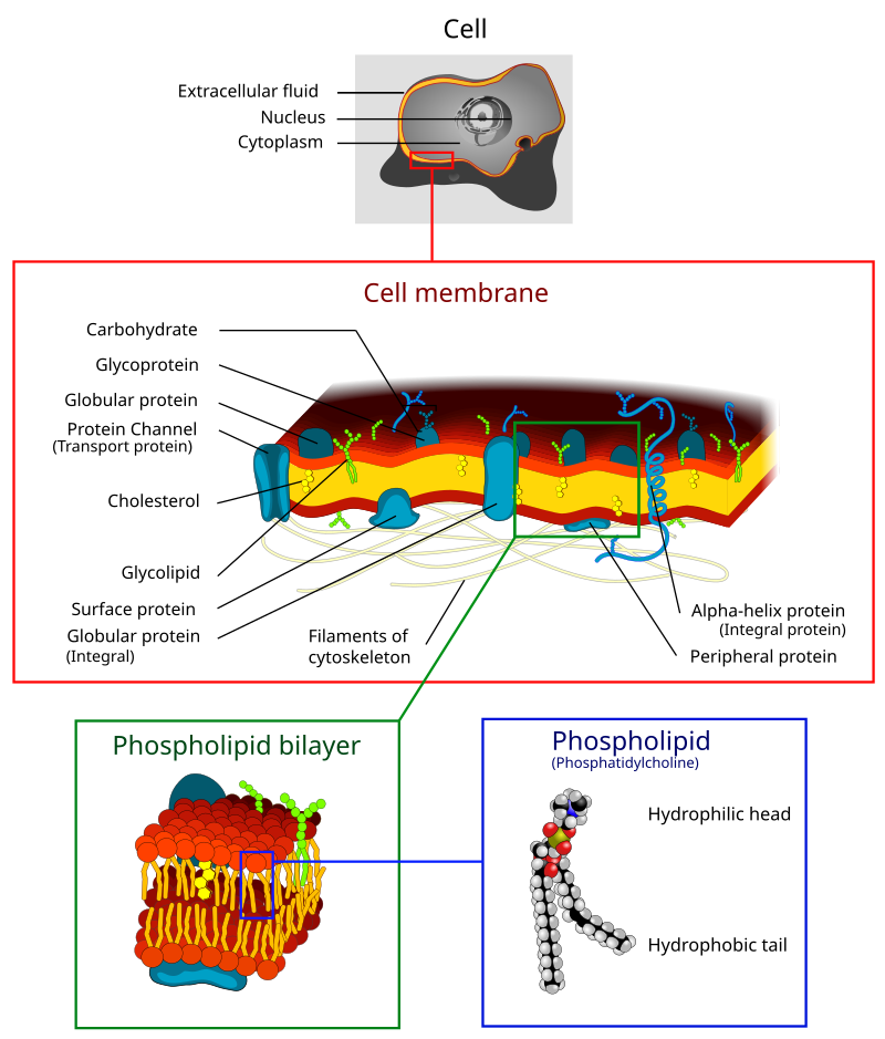
plasma membrane
biological membrane that separates the interior of a cell from its outside environment

endoplasmic reticulum
irregular network of membranes coterminous with the outer nuclear membrane in eukaryote cytoplasm that form a meshwork of tubular channels, often expanded into cisternae

Golgi apparatus
cell organelle that packages proteins for export
lysosome
A lysosome () is a membrane-bound organelle that is found in all animal cells (except red blood cells), and rarely in plant cells. There are normally hundreds of lysosomes in the cytosol, where they function as the cell's degradation center. Their primary responsibility is for catabolic degradation of proteins, polysaccharides and lipids into their respective building-block molecules: amino acids, monosaccharides, and free fatty acids. The breakdown is done by various enzymes, for example proteases, glycosidases and lipases.
cell wall
rigid or semi-rigid envelope lying outside the cell membrane of plant, fungal, most prokaryotic cells and some protozoan parasites, maintaining their shape and protecting them from osmotic lysi

nucleolus
thumb |292px |Nucleolus contained within the cell nucleus

plastid
thumb|Plant cells with visible chloroplasts

peroxisome
300px|right|thumb|Basic structure of a peroxisome
thumb|300px|Distribution of peroxisomes (white) in HEK 293 cells during [[mitosis]]
alt=Peroxisome in rat neonatal cardiomyocyte staining The SelectFX Alexa Fluor 488 Peroxisome Labeling Kit directed against peroxisomal membrane protein 70 (PMP 70)|thumb|Peroxisome in rat neonatal cardiomyocyte

flagellum
A flagellum (; : flagella) (Latin for 'whip' or 'scourge') is a hair-like appendage that protrudes from certain plant and animal sperm cells, from fungal spores (zoospores), and from a wide range of microorganisms to provide motility. Many protists with flagella are known as flagellates.

vesicle
any small, fluid-filled, spherical organelle enclosed by a membrane

cilium
The cilium (: cilia; ; in Medieval Latin and in anatomy, cilium) is a short hair-like membrane protrusion from many types of eukaryotic cell. (Cilia are absent in bacteria and archaea.) The cilium has the shape of a slender threadlike projection that extends from the surface of the much larger cell body. Eukaryotic flagella found on sperm cells and many protozoans have a similar structure to motile cilia that enables swimming through liquids, but they are longer than cilia and have a different undulating motion.

proteasome complex
thumb|right|Cartoon representation of a proteasome. Its active sites are sheltered inside the tube (blue). The caps (red; in this case, 11S regulatory particles) on the ends regulate entry into the destruction chamber, where the protein is degraded.
thumb|right|Top view of the proteasome above.
Proteasomes are essential protein complexes responsible for the degradation of proteins by proteolysis, a chemical reaction that breaks peptide bonds. Enzymes that help such reactions are called proteases. Proteasomes are found inside all eukaryotes and archaea, and in some bacteria.
In eukaryotes, prot

myofibril
A myofibril (also known as a muscle fibril or sarcostyle) is a basic rod-like organelle of a muscle cell. Skeletal muscles are composed of long, tubular cells known as muscle fibers, and these cells contain many chains of myofibrils. Each myofibril has a diameter of 1–2 micrometres. They are created during embryonic development in a process known as myogenesis.

leucoplast
thumb|250px|Leucoplasts, specifically, amyloplasts
Leucoplasts ("formed, molded") are a category of plastid and as such are organelles found in plant cells. They are non-pigmented, in contrast to other plastids such as the chloroplast.

plasmodesma
thumb|upright=1.2|The structure of a primary plasmodesma. CW=cell wall, CA=[[callose, PM=plasma membrane, ER=endoplasmic reticulum, DM=desmotubule, Red circles=actin, Purple circles and spokes=other unidentified proteins]]
amyloplast
right|thumb|Amyloplasts in a potato cellAmyloplasts are a type of plastid, double-enveloped organelles in plant cells that are involved in various biological pathways. Amyloplasts are specifically a type of leucoplast, a subcategory for colorless, non-pigment-containing plastids. Amyloplasts are found in roots and storage tissues, and they store and synthesize starch for the plant through the polymerization of glucose. Starch synthesis relies on the transportation of carbon from the cytosol, the mechanism by which is currently under debate.

spliceosome
A spliceosome is a large ribonucleoprotein (RNP) complex found primarily within the nucleus of eukaryotic cells. The spliceosome is assembled from small nuclear RNAs (snRNA) and numerous proteins. Small nuclear RNA (snRNA) molecules bind to specific proteins to form a small nuclear ribonucleoprotein complex (snRNP, pronounced "snurps"), which in turn combines with other snRNPs to form a large ribonucleoprotein complex called a spliceosome. The spliceosome removes introns from a transcribed pre-mRNA, a type of primary transcript. This process is generally referred to as splicing. An analogy is
endosome
thumb|315px|alt=endocytic pathway compartments|Electron micrograph of endosomes in human HeLa cells. Early endosomes (E - labeled for EGFR, 5 minutes after internalisation, and transferrin), late endosomes/MVBs (M) and lysosomes (L) are visible. Bar, 500 nm.

pilus
right|thumb|350px|Schematic drawing of bacterial conjugation. 1- Donor cell produces pilus. 2- Pilus attaches to recipient cell, brings the two cells together. 3- The mobile plasmid is nicked and a single strand of DNA is then transferred to the recipient cell. 4- Both cells recircularize their plasmids, synthesize second strands, and reproduce pili; both cells are now viable donors.
A pilus (Latin for 'hair'; : pili) is a hair-like cell-surface appendage found on many bacteria and archaea. The terms pilus and fimbria (Latin for 'fringe'; plural: fimbriae) can be used interchangeably, although

kinetochore
300px|thumb|Image of kinetochores in pink
melanosome
thumb|7× speed timelapse video of fish melanophores responding to 200 uM adrenaline; the melanosomes retreat to the center of the star-shaped melanophore cells.
thumb|Fish and frog melanophores are cells that can change colour by dispersing or aggregating pigment-containing melanosomes.

Mesosome
thumb|right|250px|Mesosomes form in bacterial cells prepared for electron microscopy by chemical fixation, but not by freeze-fracture fixation.
glyoxysome
Glyoxysomes are specialized peroxisomes found in plants (particularly in the fat storage tissues of germinating seeds) and also in filamentous fungi. Seeds that contain fats and oils include corn, soybean, sunflower, peanut and pumpkin. As in all peroxisomes, in glyoxysomes the fatty acids are oxidized to acetyl-CoA by peroxisomal β-oxidation enzymes. When the fatty acids are oxidized hydrogen peroxide (H2O2) is produced as oxygen (O2) is consumed. Thus the seeds need oxygen to germinate. Besides peroxisomal functions, glyoxysomes possess additionally the key enzymes of the glyoxylate cycle (i

axoneme
thumb|Electron micrograph of a thin cross-section through two Chlamydomonas axonemes
thumb|A simplified model of intraflagellar transport.

phagosome
300px|thumb | Phagocytosis of a bacterium, showing the formation of phagosome and phagolysosome
basal body
cell protein structure found at the base of cilium or flagellum

hydrogenosome
thumb|594x594px|Activity in a Spironucleus salmonicida|Spironucleus salmonicida hydrogenosome: [[pyruvate (PYR) is turned into carbon dioxide (CO2) and acetate while producing molecular hydrogen (H2) and converting ADP into ATP]]

etioplast
thumb|Different types of plastid
Etioplasts are an intermediate type of plastid that develop from proplastids that have not been exposed to light, and convert into chloroplasts upon exposure to light. They are usually found in stem and leaf tissue of flowering plants (Angiosperms) grown either in complete darkness, or in extremely low-light conditions.

cytostome
thumbnail|Diagram of a ciliate

magnetosome
[[File:Magnetite magnetosomes in Gammaproteobacteria.png |thumb |Magnetite magnetosomes in Gammaproteobacteria strain SS-5. (A) Chain of highly elongated magnetosomes. (B) Part of a magnetosome chain. (C) The magnetosome in the lower right in (B), viewed along the \scriptstyle [1\overline{1}0] direction, with its Fourier transform in the lower right.]]
Magnetosomes are membranous structures present in magnetotactic bacteria (MTB). They contain iron-rich magnetic particles that are enclosed within a lipid bilayer membrane. Each magnetosome can often contain 15 to 20 magnetite crystals that form

carboxysome
right|thumb|460px|Electron micrographs showing alpha-carboxysomes from the chemoautotrophic bacterium Halothiobacillus|Halothiobacillus neapolitanus: (A) arranged within the cell, and (B) intact upon isolation. Scale bars indicate 100 nm.
contractile vacuole
specialized vacuole of eukaryotic cells, especially Protozoa, that fills with water from the cytoplasm and then discharges this externally by the opening of contractile vacuole pores. Its function is probably osmoregulatory
kinetoplast
thumb|Electron micrograph of normal kinetoplast (K) of Trypanosoma brucei
A kinetoplast is a network of circular DNA (called kDNA) inside a mitochondrion that contains many copies of the mitochondrial genome. The most common kinetoplast structure is a disk, but they have been observed in other arrangements. Kinetoplasts are only found in Excavata of the class Kinetoplastida. The variation in the structures of kinetoplasts may reflect phylogenic relationships between kinetoplastids. A kinetoplast is usually adjacent to the organism's flagellar basal body, suggesting that it is bound to some com
mitosome
A mitosome (also called a crypton in early literature) is a mitochondrion-related organelle (MRO) found in a variety of parasitic unicellular eukaryotes, such as members of the supergroup Excavata. The mitosome was first discovered in 1999 in Entamoeba histolytica, an intestinal parasite of humans, and mitosomes have also been identified in several species of Microsporidia and in Giardia intestinalis.
Cajal body
class of nuclear body enriched in small nuclear ribonucleoproteins
sarcoplasmic reticulum
fine reticular network of membrane-limited elements that pervades the sarcoplasm of a muscle cell
microbody
A microbody (or cytosome) is a type of organelle that is found in the cells of plants, protozoa, and animals. Organelles in the microbody family include peroxisomes, glyoxysomes, glycosomes and hydrogenosomes. In vertebrates, microbodies are especially prevalent in the liver and kidney. Many membrane bound vesicles called microbodies that contain various enzymes, are present in both plant and animal cells.

Parenthesome
thumb|Diagram of situation of the fungal organelle parenthesome in the cell
Within the cells of some members of basidiomycetes fungi are found microscopic structures called parenthesomes or septal pore caps. They are shaped like parentheses and found on either side of pores in the dolipore septum which separates cells within a hypha. Their function has not been established, and their composition has not been fully elucidated. The variations in their appearance are useful in distinguishing individual species.
Generally, they are barrel shaped, with an endoplasmic reticulum covering.

nucleomorph
thumb|Diagram of a four membraned plastid containing a nucleomorph
Nucleomorphs are small, vestigial eukaryotic nuclei found between the inner and outer pairs of membranes in certain plastids. They are thought to be vestiges of red and green algal nuclei that were engulfed by a larger eukaryote. Because the nucleomorph lies between two sets of membranes, nucleomorphs support the endosymbiotic theory and are evidence that the plastids containing them are complex plastids. Having two sets of membranes indicate that the plastid, a prokaryote, was engulfed by a eukaryote, an alga, which was then e
Weibel-Palade body
large, elongated, rod-shaped secretory granule characteristic of vascular endothelial cells that contain a number of structurally and functionally distinct proteins, of which the best characterized are von Willebrand factor (VWF) and P-selectin
phycobilisome
Phycobilisomes are light-harvesting antennae that transmit the energy of harvested photons to photosystem II and photosystem I in cyanobacteria and in the chloroplasts of red algae and glaucophytes. They were lost during the evolution of the chloroplasts of green algae and plants.
vault
eukaryotic organelle with 39-fold symmetry
fluid mosaic model
commonly used model to study the structure of cell membrane
apicoplast
An apicoplast is a derived non-photosynthetic plastid found in most Apicomplexa, including Toxoplasma gondii, and Plasmodium falciparum and other Plasmodium spp. (parasites causing malaria), but not in others such as Cryptosporidium. It originated from algae through secondary endosymbiosis; there is debate as to whether this was a green or red alga. The apicoplast is surrounded by four membranes within the outermost part of the endomembrane system. The apicoplast hosts important metabolic pathways like fatty acid synthesis, isoprenoid precursor synthesis and parts of the heme biosynthetic path
type III protein secretion system complex
complex that carries out protein secretion in the bacterial type III secretion system
.png)
autophagosome
thumb|The autophagic process is divided into five distinct stages: Initiation, phagophore nucleation, autophagosomal formation (elongation), autophagosome-lysosome fusion (autophagolysosome) and cargo degradation.
An autophagosome is a spherical structure with double layer membranes. It is the key structure in macroautophagy, the intracellular degradation system for cytoplasmic contents (e.g., abnormal intracellular proteins, excess or damaged organelles, invading microorganisms). After formation, autophagosomes deliver cytoplasmic components to the lysosomes. The outer membrane of an autophag

apoptosome
thumb|alt=3D structure of the human apoptosome-CARD complex.|3D structure of the human apoptosome-CARD complex. blue: apoptosome platform; magenta: CARD disk
gerontoplast
A gerontoplast is a type of organelle known as a plastid, which develops from a chloroplast during the senescing of plant foliage. Gerontoplast development is generally seen to be the process of grana being unstacked, loss of thylakoid membranes, and large accumulation of plastoglobuli.
Tannosome
Tannosomes are organelles found in plant cells of vascular plants.

trichocyst
thumb|Paramecium tetraurelia, a ciliate, with discharged trichocysts (artificially colored in blue).
A trichocyst is an organelle found in certain ciliates and dinoflagellates.

phagolysosome
In biology, a phagolysosome, or endolysosome, is a cytoplasmic body formed by the fusion of a phagosome with a lysosome in a process that occurs during phagocytosis. Formation of phagolysosomes is essential for the intracellular destruction of microorganisms and pathogens. It takes place when the phagosome's and lysosome's membranes 'collide', at which point the lysosomal contents—including hydrolytic enzymes—are discharged into the phagosome in an explosive manner and digest the particles that the phagosome had ingested. Some products of the digestion are useful materials and are moved into t

Glycosome
The glycosome is a membrane-enclosed organelle that contains the glycolytic enzymes. The term was first used by Scott and Still in 1968 after they realized that the glycogen in the cell was not static but rather a dynamic molecule. It is found in a few species of protozoa including the Kinetoplastida which include the suborders Trypanosomatida and Bodonina, most notably in the human pathogenic trypanosomes, which can cause sleeping sickness, Chagas's disease, and leishmaniasis. The organelle is bounded by a single membrane and contains a dense proteinaceous matrix. It is believed to have evolv
eyespot apparatus
Small pigmented organelle used in single-celled organisms to detect light
extrusome
Extrusomes are membrane-bound organelles found in eukaryotic cells that are capable of discharging material contained within to the exterior of the cell. Due to the diversity in structure and function, it is unlikely that different types of extrusomes are homologous.
matrix
material or tissue between animal or plant cells
Bacterial microcompartment
organelle-like structure in bacteria with a protein shell containing enzymes

micronucleus
thumb|Micronuclei visible in boxes|280x280px
hemifusome
thumb|The green and orange structures in this image are hemifusomes, newly discovered organelles that may represent a previously unrecognized pathway for recycling in human cells.
Hemifusomes are intracellular organelles first described in 2025 by researchers at the National Institutes of Health and the University of Virginia School of Medicine. Discovered using in situ cryo-electron tomography, hemifusomes are heterotypic vesicular complexes consisting of a smaller translucent vesicle and a larger granular vesicle connected by an extended hemifusion diaphragm (HD)—a shared bilayer structure t