Category
page 1Cell anatomy

cell
basic structural and functional unit of all organisms

nucleus
membrane-bounded organelle of eukaryotic cells in which chromosomes are housed and replicated

cytoplasm
The cytoplasm is all the material within a eukaryotic or prokaryotic cell, enclosed by the cell membrane, including the organelles and excluding the nucleus in eukaryotic cells. The material inside the nucleus of a eukaryotic cell and contained within the nuclear membrane is termed the nucleoplasm. The main components of the cytoplasm are the cytosol (a gel-like substance), the cell's internal sub-structures, and various cytoplasmic inclusions. The cytoplasm is about 80% water and is usually colorless.
organelle
An organelle is a specialized subunit, within a biological cell, that has a specific function. The name organelle comes from the idea that these structures are parts of cells, as organs are to the body, hence organelle, the suffix -elle being a diminutive. Organelles are either separately enclosed within their own lipid bilayers (also called membrane-bound organelles) or are spatially distinct functional units without a surrounding lipid bilayer (non-membrane bounded organelles). Although most organelles are functional units within cells, some functional units that extend outside of cells are
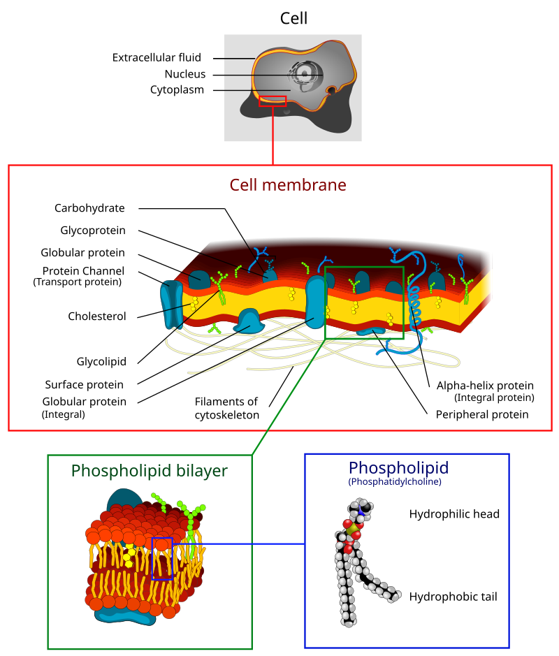
plasma membrane
biological membrane that separates the interior of a cell from its outside environment

endoplasmic reticulum
irregular network of membranes coterminous with the outer nuclear membrane in eukaryote cytoplasm that form a meshwork of tubular channels, often expanded into cisternae

vacuole
thumb|300x300px|Plant cell structure
thumb|300x300px|Animal cell structure
A vacuole () is a membrane-bound organelle which is present in plant and fungal cells and some protist, animal, and bacterial cells. Vacuoles are essentially enclosed compartments which are filled with water containing inorganic and organic molecules including enzymes in solution, though in certain cases they may contain solids which have been engulfed. Vacuoles are formed by the fusion of multiple membrane vesicles and are effectively just larger forms of these. The organelle has no basic shape or size; its structure v
lysosome
A lysosome () is a membrane-bound organelle that is found in all animal cells (except red blood cells), and rarely in plant cells. There are normally hundreds of lysosomes in the cytosol, where they function as the cell's degradation center. Their primary responsibility is for catabolic degradation of proteins, polysaccharides and lipids into their respective building-block molecules: amino acids, monosaccharides, and free fatty acids. The breakdown is done by various enzymes, for example proteases, glycosidases and lipases.

cytoskeleton
thumb|right|300px|The cytoskeleton consists of (a) microtubules, (b) microfilaments, and (c) intermediate filaments.

cytosol
thumb|right|300px|The cytosol is a crowded solution of many different types of molecules that occupy up to 30% of the cytoplasmic volume.

microtubule
alt=Tubulin and Microtubule Metrics Infographic|thumb|

endocytosis
thumb|right|400px|The different types of endocytosis
Endocytosis is a cellular process in which substances are brought into the cell. The material to be internalized is surrounded by an area of cell membrane, which then buds off inside the cell to form a vesicle containing the ingested materials. Endocytosis includes pinocytosis (cell drinking) and phagocytosis (cell eating). It is a form of active transport.

nucleoplasm
thumb|300px|The protoplasmic material of the nucleus including the [[nucleolus labelled as nucleoplasm.]]
The nucleoplasm, also known as karyoplasm, is the type of protoplasm that makes up the cell nucleus, the most prominent organelle of the eukaryotic cell. It is enclosed by the nuclear envelope, also known as the nuclear membrane. The nucleoplasm resembles the cytoplasm of a eukaryotic cell in that it is a gel-like substance found within a membrane, although the nucleoplasm only fills out the space in the nucleus and has its own unique functions. The nucleoplasm suspends structures within t

pseudopodium
thumb|Amoeba proteus extending lobose pseudopodia|300x300px

plasmodesma
thumb|upright=1.2|The structure of a primary plasmodesma. CW=cell wall, CA=[[callose, PM=plasma membrane, ER=endoplasmic reticulum, DM=desmotubule, Red circles=actin, Purple circles and spokes=other unidentified proteins]]

sarcomere
A sarcomere (Greek σάρξ sarx "flesh", μέρος meros "part") is the smallest functional unit of striated muscle tissue. It is the repeating unit between two Z-lines. Skeletal muscles are composed of tubular muscle cells (called muscle fibers or myofibers) which are formed during embryonic myogenesis. Muscle fibers contain numerous tubular myofibrils. Myofibrils are composed of repeating sections of sarcomeres, which appear under the microscope as alternating dark and light bands. Sarcomeres are composed of long, fibrous proteins as filaments that slide past each other when a muscle contracts or r
cell junction
a cellular component that forms a specialized region of connection between two or more cells, or between a cell and the extracellular matrix, or between two membrane-bound components of a cell, such as flagella
desmosome
A desmosome (; "binding body"), also known as a macula adherens (plural: maculae adherentes) (Latin for adhering spot), is a cell structure specialized for cell-to-cell adhesion. A type of junctional complex, they are localized spot-like adhesions randomly arranged on the lateral sides of plasma membranes. Desmosomes are one of the stronger cell-to-cell adhesion types and are found in tissue that experience intense mechanical stress, such as cardiac muscle tissue, bladder tissue, gastrointestinal mucosa, and epithelia.

amyloplast
right|thumb|Amyloplasts in a potato cellAmyloplasts are a type of plastid, double-enveloped organelles in plant cells that are involved in various biological pathways. Amyloplasts are specifically a type of leucoplast, a subcategory for colorless, non-pigment-containing plastids. Amyloplasts are found in roots and storage tissues, and they store and synthesize starch for the plant through the polymerization of glucose. Starch synthesis relies on the transportation of carbon from the cytosol, the mechanism by which is currently under debate.
spindle
array of microtubules and associated molecules that forms between opposite poles of a eukaryotic cell during mitosis or meiosis and serves to move the duplicated chromosomes apart
gap junction
cell-cell junction comprised of innexins or connexins, two different families of channel-forming proteins. Other proteins may also be present.
endomembrane system
collection of membranous structures involved in transport within the cell. The main components of the endomembrane system are endoplasmic reticulum, Golgi bodies, vesicles, cell membrane and nuclear envelope
tight junctions
type of cellular connection that minimizes metabolic flows between adjacent cells
plastome
DNA located in cellular organelles called plastids
hemidesmosome
Hemidesmosomes are very small stud-like structures found in keratinocytes of the epidermis of skin that attach to the extracellular matrix. They are similar in form to desmosomes when visualized by electron microscopy; however, desmosomes attach to adjacent cells. Hemidesmosomes are also comparable to focal adhesions, as they both attach cells to the extracellular matrix. Instead of desmogleins and desmocollins in the extracellular space, hemidesmosomes utilize integrins. Hemidesmosomes are found in epithelial cells connecting the basal epithelial cells to the basement membrane. Hemidesmosomes

phagosome
300px|thumb | Phagocytosis of a bacterium, showing the formation of phagosome and phagolysosome
Acinus
An acinus (; : acini; adjective, acinar or acinous) refers to any cluster of cells that resembles a many-lobed "berry", such as a raspberry (acinus is Latin for "berry"). The berry-shaped termination of an exocrine gland, where the secretion is produced, is acinar in form, as is the alveolar sac containing multiple alveoli in the lungs.
adherens junction
cell junction at which anchoring proteins extend through the membrane and attach to actin filaments
ectoplasm
part of a cell's cytoplasm
neurite
A neurite or neuronal process refers to any projection from the cell body of a neuron. This projection can be either an axon or a dendrite. The term is frequently used when speaking of immature or developing neurons, especially of cells in culture, because it can be difficult to tell axons from dendrites before differentiation is complete.

S-layer
An S-layer (surface layer) is a part of the cell envelope found in almost all archaea, as well as in many types of bacteria.
The S-layers of both archaea and bacteria consists of a monomolecular layer composed of only one (or, in a few cases, two) identical proteins or glycoproteins. This structure is built via self-assembly and encloses the whole cell surface. Thus, the S-layer protein can represent up to 15% of the whole protein content of a cell. S-layer proteins are poorly conserved or not conserved at all, and can differ markedly even between related species. Depending on species, the S-l
sarcoplasm
Sarcoplasm is the cytoplasm of a muscle cell. It is comparable to the cytoplasm of other cells, but it contains unusually large amounts of glycogen (a polymer of glucose), myoglobin, a red-colored protein necessary for binding oxygen molecules that diffuse into muscle fibers, and mitochondria. The calcium ion concentration in sarcoplasm is also a special element of the muscle fiber; it is the means by which muscle contractions take place and are regulated. The sarcoplasm plays a critical role in muscle contraction as an increase in concentration in the sarcoplasm begins the process of filament
T-tubule
T-tubules (transverse tubules) are extensions of the cell membrane that penetrate into the center of skeletal and cardiac muscle cells. With membranes that contain large concentrations of ion channels, transporters, and pumps, T-tubules permit rapid transmission of the action potential into the cell, and also play an important role in regulating cellular calcium concentration.
Granule
part of a cell
mitochondrial matrix
The gel-like material, with considerable fine structure, that lies in the matrix space, or lumen, of a mitochondrion. It contains the enzymes of the tricarboxylic acid cycle and, in some organisms, the enzymes concerned with fatty acid oxidation.

carboxysome
right|thumb|460px|Electron micrographs showing alpha-carboxysomes from the chemoautotrophic bacterium Halothiobacillus|Halothiobacillus neapolitanus: (A) arranged within the cell, and (B) intact upon isolation. Scale bars indicate 100 nm.

ultrastructure
thumb|right|The ultrastructure of a single Bacterium|bacterial cell ([[Bacillus subtilis). The scale bar is 200 nm.]]
Cajal body
class of nuclear body enriched in small nuclear ribonucleoproteins
caveola
In biology, caveolae (Latin for "little caves"; singular, caveola), which are a special type of lipid raft, are small (50–100 nanometer) invaginations of the plasma membrane in the cells of many vertebrates. They are the most abundant surface feature of many vertebrate cell types, especially endothelial cells, adipocytes and embryonic notochord cells. They were originally discovered by E. Yamada in 1955.
phycobilisome
Phycobilisomes are light-harvesting antennae that transmit the energy of harvested photons to photosystem II and photosystem I in cyanobacteria and in the chloroplasts of red algae and glaucophytes. They were lost during the evolution of the chloroplasts of green algae and plants.
endoplasm
thumb|Shown is a micrograph of an amoeba; the darker pink nucleus is central to the eukaryotic cell, with the majority of the rest of the cell's body belonging to the endoplasm. Though not visible, the ectoplasm resides directly internal to the plasma membrane.
Endoplasm, also known as entoplasm, generally refers to the inner (often granulated), dense part of a cell's cytoplasm. The nucleus is separated from the endoplasm by the nuclear envelope. In an amoeba and other protists the outer part of the cytoplasm is known as the ectoplasm. The different makeups/viscosities of the endoplasm and ect
fluid mosaic model
commonly used model to study the structure of cell membrane
Prokaryotic cytoskeleton
structural filaments in prokaryotes
.png)
autophagosome
thumb|The autophagic process is divided into five distinct stages: Initiation, phagophore nucleation, autophagosomal formation (elongation), autophagosome-lysosome fusion (autophagolysosome) and cargo degradation.
An autophagosome is a spherical structure with double layer membranes. It is the key structure in macroautophagy, the intracellular degradation system for cytoplasmic contents (e.g., abnormal intracellular proteins, excess or damaged organelles, invading microorganisms). After formation, autophagosomes deliver cytoplasmic components to the lysosomes. The outer membrane of an autophag
epiretinal membrane
disease of the eye in response to changes in the vitreous humor or more rarely, diabetes
cellular compartment
closed part in cytosol

Cystolith
thumb|Cystolith from leaf of Ficus elastica
thumb|Drawing of a cystolith from leaf of Ficus elastica
nucleolus organizer region
region of a chromosome where nucleoli form during interphase, and where genes encoding the largest rRNA precursor transcript are tandemly arrayed
matrix
material or tissue between animal or plant cells
intermembrane space
part of a cell
oil body
lipid-containing structure found in plant cells
Hormogonium
thumb|Nostoc with hormogonia
Hormogonia are motile filaments of cells formed by some cyanobacteria in the order Nostocales and Stigonematales. They are formed during vegetative reproduction in unicellular, filamentous cyanobacteria, and some may contain heterocysts and akinetes.
Phragmosome
thumb|Phragmosome formation in a highly vacuolated plant cell. From top to bottom: 1) Interphase cell with large central vacuole. 2) Cytoplasmic strands starting to penetrate vacuole. 3) Nucleus migration into center and formation of the phragmosome. 4) Phragmosome formation completed and formation of preprophase band marking future cell division plane.
tunneling nanotube
group of membrane proteins facilitating the intercellular transport of various cellular components
volutin granules
complexed inorganic polyphosphate
Porosome
thumb|440px
thumb|280px Porosomes are cup-shaped supramolecular structures in the cell membranes of eukaryotic cells where secretory vesicles transiently dock in the process of vesicle fusion and secretion. The transient fusion of secretory vesicle membrane at a porosome, base via SNARE proteins, results in the formation of a fusion pore or continuity for the release of intravesicular contents from the cell. After secretion is complete, the fusion pore temporarily formed at the base of the porosome is sealed. Porosomes are few nanometers in size and contain many different types of protein, es
undulipodium
An undulipodium or undulopodium (Greek: "swinging foot"; plural undulipodia) is a motile filamentous extension of eukaryotic cells, composed of a membrane protrusion held by a cytoskeletal structure called the axoneme. It is divided into cilia and flagella – which are differing terms for structurally identical organelles used on different types of cells, but are distinguished according to function and/or length, and usually corresponds to different waveforms of the organelles beating motion. The Gene Ontology database does not make a distinction between the two, referring to most undulipodia a
paraspeckle
frame|An overlay of a fluorescence micrograph (green) onto a DIC image of a HeLa cell expressing a Yellow fluorescent Protein fusion of Paraspeckle Protein 1 (PSP1): 1. cytoplasm; 2. nucleus; 3. nucleolus; 4. paraspeckles
In cell biology, a paraspeckle is an irregularly shaped compartment of the cell, approximately 0.2-1 μm in size, found in the nucleus' interchromatin space. First documented in HeLa cells, where there are generally 10-30 per nucleus, Paraspeckles are now known to also exist in all human primary cells, transformed cell lines and tissue sections. Their name is derived from thei
secondary cell wall
plant cell wall that is no longer able to expand and so does not permit growth. Secondary cell walls contain less pectin than that of primary cell walls. The secondary cell is mostly composed of cellulose and is strengthened with lignin
Stromule
A stromule is a microscopic structure found in plant cells. Stromules (stroma-filled tubules) are highly dynamic structures extending from the surface of all plastid types, including proplastids, chloroplasts, etioplasts, leucoplasts, amyloplasts, and chromoplasts. Protrusions from and interconnections between plastids were observed in 1888 (Gottlieb Haberlandt) and 1908 (Gustav Senn) and have been described sporadically in the literature since then. Stromules were recently rediscovered in 1997 and have since been reported to exist in a number of angiosperm species including Arabidopsis thalia