Category
page 1Membrane biology
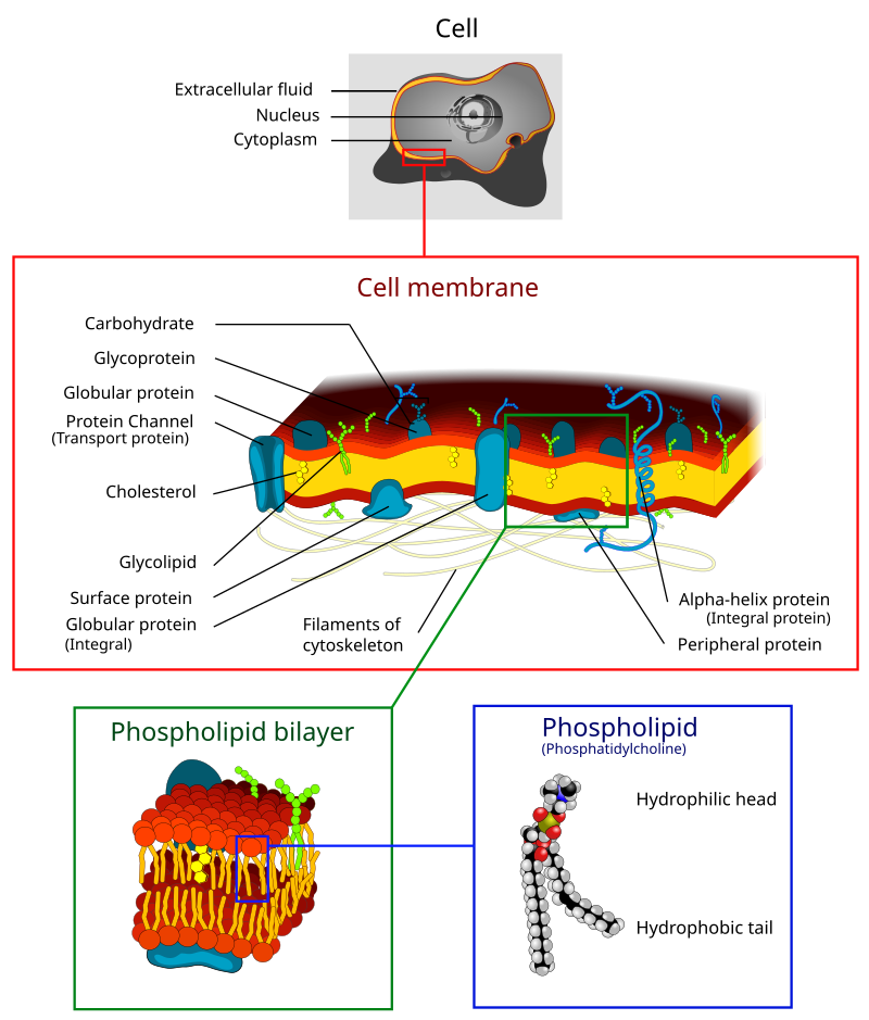
plasma membrane
biological membrane that separates the interior of a cell from its outside environment

action potential
process by which neurons communicate with each other by changes in their membrane potentials.

mucous membrane
protective layer which lines the interior of hollow organs
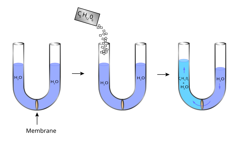
osmotic pressure
measure of the tendency of a solution to take in pure solvent by osmosis

vesicle
any small, fluid-filled, spherical organelle enclosed by a membrane

signaling receptor
protein molecule receiving signals for a cell

nuclear membrane
membrane part of the nuclear envelope

endocytosis
thumb|right|400px|The different types of endocytosis
Endocytosis is a cellular process in which substances are brought into the cell. The material to be internalized is surrounded by an area of cell membrane, which then buds off inside the cell to form a vesicle containing the ingested materials. Endocytosis includes pinocytosis (cell drinking) and phagocytosis (cell eating). It is a form of active transport.

peptidoglycan
Peptidoglycan, murein or mucopeptide is a unique large macromolecule, a polysaccharide, consisting of sugars and amino acids that forms a mesh-like layer (sacculus) that surrounds the bacterial cytoplasmic membrane. The sugar component consists of alternating residues of β-(1,4) linked N-acetylglucosamine (NAG) and N-acetylmuramic acid (NAM). Attached to the N-acetylmuramic acid is an oligopeptide chain made of three to five amino acids. The peptide chain can be cross-linked to the peptide chain of another strand forming the 3D mesh-like layer. Peptidoglycan serves a structural role in the bac
lipid bilayer
thin polar membrane made of two layers of lipid molecules

active transport
cellular transport mechanism

exocytosis
thumb|upright=1.35|Exocytosis of neurotransmitters into a synapse from neuron A to neuron B.
Exocytosis () is a form of active transport in which a cell transports molecules (e.g., neurotransmitters and proteins) out of the cell. As an active transport mechanism, exocytosis requires the use of energy to transport material. Exocytosis and its counterpart, endocytosis, are used by all cells because most chemical substances important to them are large polar molecules that cannot pass through the hydrophobic portion of the cell membrane by passive means. Exocytosis is the process by which a large

biofilm
thumb|right|300px|Staphylococcus aureus biofilm on an indwelling [[catheter]]

micelles
thumb|250px|right|Cross-section view of the structures that can be formed by phospholipids in aqueous solutions (unlike this illustration, micelles are usually formed by single-chain lipids, since it is difficult to fit two chains into this shape)
thumb|250px|right|Scheme of a micelle formed by phospholipids in an [[aqueous solution]]

amnion
The amnion (: amnions or amnia) is a membrane that closely covers human and various other embryos when they first form. It fills with amniotic fluid, which causes the amnion to expand and become the amniotic sac that provides a protective environment for the developing embryo. The amnion, along with the chorion, the yolk sac and the allantois protect the embryo. In birds, reptiles and monotremes, the protective sac is enclosed in a shell. In marsupials and placental mammals, it is enclosed in a uterus.
semipermeable membrane
membrane which will allow certain molecules or ions to pass through it by diffusion

protoplast
thumb|right|Protoplasts of cells from a petunia's leaf
thumb|Protoplasts of the moss Physcomitrella patens
Protoplast (), is a biological term coined by Hanstein in 1880 to refer to the entire cell, excluding the cell wall. Protoplasts can be generated by stripping the cell wall from plant, bacterial, or fungal cells by mechanical, chemical or enzymatic means.
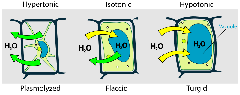
turgor pressure
intracellular pressure exerted by the plasma against the cell wall

thylakoid
250px|thumb|Thylakoids (dark green) inside a chloroplast
Thylakoids are membrane-bound compartments inside chloroplasts and cyanobacteria. They are the site of the light-dependent reactions of photosynthesis. Thylakoids consist of a thylakoid membrane surrounding a thylakoid lumen. Chloroplast thylakoids frequently form stacks of disks referred to as grana (singular: granum). Grana are connected by intergranal or stromal thylakoids, which join granum stacks together as a single functional compartment.
biological membrane
selectively permeable membrane that separates the interior of a cell from the external environment or creates intracellular compartments by serving as a boundary between one part of the cell and another

plasmolysis
Plasmolysis is the process in which cells lose water in a hypertonic solution. The reverse process, deplasmolysis or cytolysis, can occur if the cell is in a hypotonic solution resulting in a lower external osmotic pressure and a net flow of water into the cell. Through observation of plasmolysis and deplasmolysis, it is possible to determine the tonicity of the cell's environment as well as the rate solute molecules cross the cellular membrane.
node of Ranvier
axon part that is a gap in the myelin where voltage-gated sodium channels cluster and saltatory conduction takes place
nuclear pore
discrete opening in the nuclear envelope of a eukaryotic cell

liposome
thumb|right|Scheme of a liposome formed by phospholipids in an [[aqueous solution.]]
thumb|right|Liposomes are composite structures made of phospholipids and may contain small amounts of other molecules. Though liposomes can vary in size from low micrometer range to tens of micrometers, unilamellar liposomes, as pictured here, are typically in the lower size range with various targeting ligands attached to their surface allowing for their surface-attachment and accumulation in pathological areas for treatment of disease.

osmoregulation
Osmoregulation is the active regulation of the osmotic pressure of an organism's body fluids, detected by osmoreceptors, to maintain the homeostasis of the organism's water content; that is, it maintains the fluid balance and the concentration of electrolytes (salts in solution which in this case is represented by body fluid) to keep the body fluids from becoming too diluted or concentrated. Osmotic pressure is a measure of the tendency of water to move into one solution from another by osmosis. The higher the osmotic pressure of a solution, the more water tends to move into it. Pressure must

depolarization
In biology, depolarization or hypopolarization is a change within a cell, during which the cell undergoes a shift in electric charge distribution, resulting in less negative charge inside the cell compared to the outside. Depolarization is essential to the function of many cells, communication between cells, and the overall physiology of an organism.
thumb|right|Action potential in a [[neuron, showing depolarization, in which the cell's internal charge becomes less negative (more positive), and repolarization, where the internal charge returns to a more negative value.]]
Most cells in higher o

phosphatidyl serine
Phosphatidylserine (abbreviated Ptd-L-Ser or PS) is a phospholipid and is a component of the cell membrane. It plays a key role in cell cycle signaling, specifically in relation to apoptosis. It is a key pathway for viruses to enter cells via apoptotic mimicry. Its exposure on the outer surface of a membrane marks the cell for destruction via apoptosis.

N-acetyl-D-glucosamine (complete stereochemistry)
thumb|N-Acetylglucosamine molecule
'''N-Acetylglucosamine''' (GlcNAc) is an amide derivative of the monosaccharide glucose. It is a secondary amide between glucosamine and acetic acid. It is significant in several biological systems.
serous membrane
smooth coating of body cavities (thorax and abdomen), smooth, shiny, translucent and content with a thin layer of liquid
resting potential
The resting membrane potential is relatively stable and can be called as the ground value for transmembrane voltage.
ligand-gated ion channel
type of ion channel transmembrane protein
passive transport
membrane transport that occurs down an electrochemical gradient and does not require energy

sphingomyelin
thumb|General structures of sphingolipids|349x349px
cellular transport
collection of mechanisms that regulate the passage of solutes through biological membranes

lanosterol
Lanosterol is a tetracyclic triterpenoid and is the compound from which all animal and fungal steroids are derived. By contrast, plant steroids are produced via cycloartenol. In the eyes of vertebrates, lanosterol is a natural constituent, having a role in maintaining health of the lens. Lanosterol is the precursor to cholesterol.

Mesosome
thumb|right|250px|Mesosomes form in bacterial cells prepared for electron microscopy by chemical fixation, but not by freeze-fracture fixation.
endomembrane system
collection of membranous structures involved in transport within the cell. The main components of the endomembrane system are endoplasmic reticulum, Golgi bodies, vesicles, cell membrane and nuclear envelope

glycerophospholipid
thumb|right|upright=1.5|Membrane structures. Top, an archaeal phospholipid: 1, isoprene chains; 2, ether linkages; 3, Levorotation and dextrorotation|L-glycerol moiety; 4, phosphate group. Middle, a bacterial or eukaryotic phospholipid: 5, fatty acid chains; 6, ester linkages; 7, D-glycerol moiety; 8, phosphate group. Bottom: 9, lipid bilayer of bacteria and eukaryotes; 10, lipid monolayer of some archaea.
protobiont
A protocell (or protobiont) is a self-organized, membrane-bound or membraneless compartment that concentrates biomolecules, proposed as a rudimentary precursor to cells during the origin of life. A central question in evolution is how simple protocells first arose and how their progeny could diversify, thus enabling the accumulation of novel biological emergences over time (i.e. biological evolution). Although a functional protocell has not yet been achieved in a laboratory setting, the goal to understand the process appears well within reach.
electrochemical gradient
gradient of electrochemical potential, usually for an ion that can move across a membrane

phosphatidylinositols
Phosphatidylinositol or inositol phospholipid is a biomolecule. It was initially called "inosite" when it was discovered by Léon Maquenne and Johann Joseph von Scherer in the late 19th century. It was discovered in bacteria but later also found in eukaryotes, and was found to be a signaling molecule.
α-N-acetyl-D-galactosamine
'''N-Acetylgalactosamine (GalNAc'''), is an amino sugar derivative of galactose.

perylene
Perylene or perilene is a polycyclic aromatic hydrocarbon with the chemical formula C20H12, occurring as a brown solid. It or its derivatives may be carcinogenic, and it is considered to be a hazardous pollutant. In cell membrane cytochemistry, perylene is used as a fluorescent lipid probe. It is the parent compound of a class of rylene dyes.
phosphatidylethanolamines
thumb|250 px|Biosynthesis of various phospholipids (including phosphatidylethanolamine) in bacteria
N-acetyl-beta-muramic acid
chemical compound

mitochondrial crista
A crista (; : cristae) is a fold in the inner membrane of a mitochondrion. The name is from the Latin for crest or plume, and it gives the inner membrane its characteristic wrinkled shape, providing a large amount of surface area for chemical reactions to occur on. This aids aerobic cellular respiration, because the mitochondrion requires oxygen. Cristae are studded with proteins, including ATP synthase and a variety of cytochromes.

cardiolipin
Cardiolipin (IUPAC name '1,3-bis(sn-3’-phosphatidyl)-sn-glycerol', "sn" designating stereospecific numbering) is an important component of the inner mitochondrial membrane, where it constitutes about 20% of the total lipid composition. It can also be found in the membranes of most bacteria. The name "cardiolipin" is derived from the fact that it was first found in animal hearts. It was first isolated from the beef heart in the early 1940s by Mary C. Pangborn. In mammalian cells, but also in plant cells, cardiolipin (CL) is found almost exclusively in the inner mitochondrial membrane, where it
membrane raft
small sterol- and sphingolipid-enriched membrane domains that compartmentalize cellular processes
microsome
In cell biology, microsomes are heterogeneous vesicle-like artifacts (~20-200 nm diameter) re-formed from pieces of the endoplasmic reticulum (ER) when eukaryotic cells are broken-up in the laboratory; microsomes are not present in healthy, living cells.
sarcolemma
The sarcolemma (sarco (from sarx) from Greek; flesh, and lemma from Greek; sheath), also called the myolemma, is the cell membrane surrounding a skeletal muscle fibre or a cardiomyocyte.
It consists of a lipid bilayer and a thin outer coat of polysaccharide material (glycocalyx) that contacts the basement membrane. The basement membrane contains numerous thin collagen fibrils and specialized proteins such as laminin that provide a scaffold to which the muscle fibre can adhere. Through transmembrane proteins in the plasma membrane, the actin skeleton inside the cell is connected to the basemen
glycophosphatidylinositol
Glycosylphosphatidylinositol () or glycophosphatidylinositol (GPI) is a phosphoglyceride that can be attached to the C-terminus of a protein during posttranslational modification. The resulting GPI-anchored proteins play key roles in a wide variety of biological processes. GPI is composed of a phosphatidylinositol group linked through a carbohydrate-containing linker (glucosamine and mannose glycosidically bound to the inositol residue) and via an ethanolamine phosphate (EtNP) bridge to the C-terminal amino acid of a mature protein. The two fatty acids within the hydrophobic phosphatidyl-inosi

ionophore
thumb|right|420 px|Carrier and channel ionophores
(a) Carrier ionophores reversibly bind ions and carry them through cell membranes.
(b) Channel ionophores create channels within cell membranes to facilitate the transport of ions.

S-layer
An S-layer (surface layer) is a part of the cell envelope found in almost all archaea, as well as in many types of bacteria.
The S-layers of both archaea and bacteria consists of a monomolecular layer composed of only one (or, in a few cases, two) identical proteins or glycoproteins. This structure is built via self-assembly and encloses the whole cell surface. Thus, the S-layer protein can represent up to 15% of the whole protein content of a cell. S-layer proteins are poorly conserved or not conserved at all, and can differ markedly even between related species. Depending on species, the S-l
T-tubule
T-tubules (transverse tubules) are extensions of the cell membrane that penetrate into the center of skeletal and cardiac muscle cells. With membranes that contain large concentrations of ion channels, transporters, and pumps, T-tubules permit rapid transmission of the action potential into the cell, and also play an important role in regulating cellular calcium concentration.
bacterial outer membrane
plasma membrane found in gram-negative bacteria

prenylation
thumb|Skeletal formula of the prenyl group.Prenylation (also known as isoprenylation or lipidation) is the addition of hydrophobic molecules to a protein or a biomolecule. It is usually assumed that prenyl groups (3-methylbut-2-en-1-yl) facilitate attachment to cell membranes, similar to lipid anchors like the GPI anchor, though direct evidence of this has not been observed. Prenyl groups (also called isoprenyl groups, having one hydrogen atom more than isoprene) have been shown to be important for protein–protein binding through specialized prenyl-binding domains.

cytolysis
thumb|230px|right|Blood cells in solutions with different osmotic pressure. Cytolysis would result in the image on the far right.
thumb|250px|Micrographs of osmotic pressure on red blood cells
thumb|A human white blood cell (upper right) in water swells until it bursts (at ~14 seconds)
Cytolysis, or osmotic lysis, occurs when a cell bursts due to an osmotic imbalance that has caused excess water to diffuse into the cell. Water can enter the cell by diffusion through the cell membrane or through selective membrane channels called aquaporins, which greatly facilitate the flow of water. It occurs
hyperpolarization
change in a cell membrane potential causing it to become more negative
mycolic acids
class of compounds
L-type calcium channel
family of transport proteins